
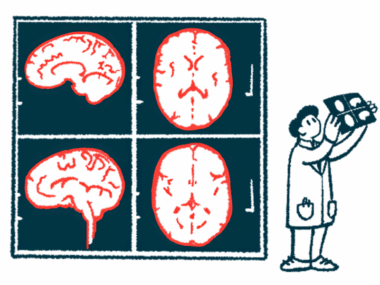
Potential biomarkers seen in protein levels in amygdala region of brain
Amygdala involved in memory and emotional responses like anxiety and fear
Written by |
Altered levels of proteins in the amygdala, the brain region involved in memory, decision-making, and emotional responses, were discovered in Parkinson’s disease patients, a study reports.
These changes in the amygdala were associated with synapses, the minute gaps between nerve cells through which impulses pass, allowing the cells to communicate.
Affected proteins “stand out as potential biomarkers in Parkinson’s disease,” the researchers wrote.

Proteins, possible biomarkers for Parkinson’s, interact with alpha-synuclein
The study, “Synaptic involvement of the human amygdala in Parkinson’s disease,” was published in the journal Molecular & Cellular Proteomics.
Alpha-synuclein is a protein primarily found in synapses at the end of nerve fibers. In people with Parkinson’s disease, the toxic buildup of alpha-synuclein clumps, known as Lewy bodies, is thought to cause cell death in a brain region called the substantia nigra, triggering the onset of Parkinson’s motor symptoms.
Studies suggest Lewy bodies also affect the amygdala in Parkinson’s. Composed of two almond-shaped clusters of nerve cells within the brain, the amygdala plays a role in memory, learning, the senses, and emotional responses such as fear, anxiety, and aggression.
Accordingly, damage to the amygdala provokes certain nonmotor disease symptoms, including anxiety, depression, fatigue, the loss of smell, and problems with the autonomic nervous system that controls unconscious processes like heart rate, digestion, and blood pressure.
Researchers in Spain analyzed proteins that interact with alpha-synuclein and other synaptic proteins in amygdala tissue taken from eight deceased Parkinson’s patients. Their goal was to identify pathways involved in the disease’s development, as well as potential therapeutic targets and biomarkers.
“Studying the role of synaptic proteins in [Parkinson’s disease] is crucial given the synaptic location of [alpha-synuclein],” the researchers wrote.
They identified 199 proteins in patients’ amygdala tissues that were produced at different levels compared with control samples from eight people who died of causes unrelated to Parkinson’s or other neurodegenerative diseases.
Of these, 25 proteins interacted with alpha-synuclein, and all of them could be found in various body fluids, including blood, saliva, urine, and cerebrospinal fluid, the liquid surrounding the brain and spinal cord. Levels of 46 synaptic proteins also differed between Parkinson’s and control samples.
Seven proteins were selected based on the criteria that they were produced at levels at least two times higher or lower than proteins in control tissues, and that they had no known relationship with Parkinson’s. Five of these proteins were at higher levels — GNAI1, EEF1A1, PLP1, NPTN, and YWHAY — and two were at lower levels — GRIM19 and ORM2.
Quantitative analysis confirmed significant increases in GNAI1, PLP1, and NPTN, and significant decreases in ORM2, in Parkinson’s amygdala samples. EEF1A1, YWHAH, and GRIM19 did not show significant differences between Parkinson’s and control group samples.
Localization tests found EEF1A1 in nerve cells, PLP1 in nerve fibers (axons), and YWHAH in the nucleus of nerve cells. ORM2 was found in astrocytes, a cell type that supports nerve cells, while NPTN and GNAI1 were dispersed throughout the tissue without specific localization. Researchers could not study the localization of GRIM19.
None of the identified proteins were found in microglia, the primary immune cells of the brain and spinal cord.
A stereological analysis then examined the shapes and sizes of cells and tissues. It found cell densities within amygdala tissue, as well as the total amygdala volume, to be similar between patient and control samples.
“Our study is the first to analyze the proteome of the [amygdala] of patients with [Parkinson’s disease] and revealed significant damage at the synaptic level,” the scientists wrote. Proteomics is a large-scale study of proteins.
This work also “identified possible biomarkers of the disease,” they added, noting that GNAI1, EEF1A1, PLP1, NPTN, YWHAH, and ORM2 can be found in fluids that include the cerebrospinal fluid.
“It seems clear that Lewy pathology is not sufficient to cause neurodegeneration or alteration of microglial and astroglial populations in the [amygdala] of humans with [Parkinson’s disease],” the team concluded.
“However, damage at the proteomic level is evident, highlighting significant synaptic involvement.”


Leave a comment
Fill in the required fields to post. Your email address will not be published.