
Stem cell therapy for Parkinson’s boosted by immune Tregs: Study
New strategy used in rats may help achieve better outcomes in people
Written by |
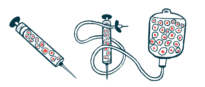
Scientists have shown that co-transplanting stem cells with regulatory T-cells — immune cells known as Tregs that dampen excessive inflammatory and immune responses — can boost cell survival in the brain in rodent models of Parkinson’s disease, and also ease motor symptoms.
These findings propose a new strategy to achieve better clinical outcomes for stem cell-based therapies for Parkinson’s in humans. Such therapies aim to replace the dopamine-producing nerve cells that are lost in people with the neurodegenerative disorder.
But to date, these stem cells have failed to thrive and survive over the long term.
“We have now made a major breakthrough using immune cells to improve delivery, survival, and recovery for neuronal cell therapies. Our findings show the power and flexibility of cell therapy to be modified and enhanced to become a realistic modality to treat conditions like Parkinson’s,” Kwang-Soo Kim, PhD, a researcher at the molecular neurobiology laboratory at McLean Hospital, in Massachusetts, and the study’s lead author, said in a press release.
The results were described in the study, “Co-transplantation of autologous Treg cells in a cell therapy for Parkinson’s disease,” published in the journal Nature.

Co-transplant aims to ease motor symptoms of Parkinson’s
The hallmark of Parkinson’s disease is the gradual decline of dopaminergic nerve cells — those responsible for producing dopamine — in a specific area of the midbrain known as the substantia nigra. Dopamine plays a crucial role as a neurotransmitter in the brain.
Existing treatments, such as levodopa, focus on restoring depleted levels of dopamine; however, these treatments only provide symptomatic relief, particularly for tremors or stiffness. Moreover, their effectiveness tends to diminish over time.
Efforts are underway to develop strategies to replace the lost dopamine-producing neurons using cell-based therapies. One of the strategies is to use midbrain dopaminergic neurons derived from stem cells, such as induced pluripotent stem cells (iPSCs) or embryonic stem cells. Both cells are able to generate almost any type of cell in the body.
However, while these cell-based therapies aim to tackle the disease’s underlying cause, efforts so far have fallen short, with the transplanted dopaminergic neurons failing to implant and survive for long.
Understanding the local reaction of brain tissue receiving the transplanted cells, particularly the response of immune cells, thus is paramount to overcome these challenges.

Rat model used to study Tregs transplanted alongside immune cells
Now, scientists at McLean and Massachusetts General Hospital have turned to a rodent model to advance their work. The team transplanted iPSCs-derived midbrain dopamine progenitor cells (mDAPs) from a patient with Parkinson’s into the brain of rodents. Dopamine progenitor cells are specialized stem cells that can become dopamine-producing nerve cells. They play a crucial role in developing and regenerating dopamine neurons in the brain.
The team had previously found that the brain surgery itself triggers an acute brain inflammation (neuroinflammation) followed by the infiltration of circulating immune cells, an adverse immune response they termed “needle-trauma.” Most importantly, analysis of the brain tissue two weeks after the transplant showed that less than 10% of the mDAPs cells had survived. The same pattern was seen when they transplanted cells from two other sources.
Given the inflammatory response observed, the researchers speculated that this could be the underlying cause of the rapid death of the transplanted cells. To address this issue, they hypothesized that co-transplanting regulatory T cells, also known as Tregs, alongside the mDAPs could potentially alleviate the acute neuroinflammation. Tregs are a type of immune cells that play a role in maintaining a balanced immune system by suppressing excessive immune responses.
The researchers used a mouse model of Parkinson’s and transplanted the mDAPs along with Tregs into the striatum — a brain region involved in voluntary movement control.
The results showed that the presence of Tregs enhanced the survival of the transplanted mDAPs, with animals showing improvements in their motor behavior.
“Initially, just one or two weeks after transplantation, the majority of the dopamine neurons died, rendering the cell therapy unsuccessful,” Kim said. “But when we added regulatory T cells to the transplant, survival of the grafted dopamine neurons increased. Also, behavior recovery was faster and more robust.”

Added benefit seen when transplanting Tregs and stem cells
Transplanting both cell types also showed an additional benefit: it suppressed the outgrowth of non-dopaminergic cells, including reactive inflammatory cells.
“This finding is very significant because a potential hazard associated with cell transplantation is often the outgrowth of undesirable, potentially harmful cells,” Kim said. “The most important criterion for cell therapy is safety.”
The results also carry significant implications for other diseases of the nervous system that might benefit from these cell-based therapies.
“Needle trauma is a universal issue in cell therapies in the nervous system, not just for dopaminergic neurons or Parkinson’s disease,” said Bob Carter, MD, PhD, chief of neurosurgery at Mass General and one of the study’s authors.
“Our principles can be applied widely to any cell therapy for other (neuro)degenerative disorders such as Alzheimer’s, ALS [amyotrophic lateral sclerosis], or Huntington’s,” Carter said.
This finding is very significant because a potential hazard associated with cell transplantation is often the outgrowth of undesirable, potentially harmful cells. The most important criterion for cell therapy is safety.
One limitation of the this study is the use of rodent models. According to Kim, future studies are needed to further assess the safety of a co-transplant, as well as to understand the mechanisms by which Tregs enhance the survival of dopaminergic neurons.
Overall, these findings suggest that the co-transplantation system described here “effectively reduces the needle trauma induced death of mDANs, providing a potential strategy to achieve better clinical outcomes for cell therapy in Parkinson’s disease,” the researchers worte.
To bring these findings closer to human clinical trials, the Mass General Brigham has launched its Gene and Cell Therapy Institute. The institute hopes to translate scientific discoveries into life-changing treatments for patients.


Leave a comment
Fill in the required fields to post. Your email address will not be published.