
Study Challenges View of How Alpha-synuclein Drives Parkinson’s
Results point to 'alternative, more complex' mechanism behind disease progression
Written by |
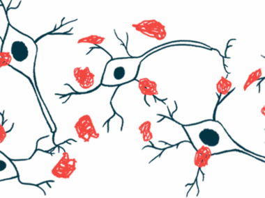
Alpha-synuclein clumps’ toxic effects — a hallmark of Parkinson’s disease — are independent of the production of the protein by neurons themselves, according to a study in mice.
In mice lacking alpha-synuclein, exposure to toxic protein clumping resulted in comparable neurodegeneration, which was found to be related to the direct toxic effects on mitochondria, the cells’ powerhouses.
The findings challenge a long-held view about how alpha-synuclein contributes to Parkinson’s, wherein toxic alpha-synuclein clumps would enter neurons and form more aggregates with normal alpha-synuclein, causing neurodegeneration.
The results point to “an alternative, more complex, molecular mechanism underlying the progression of PD [Parkinson’s] in patients,” the researchers wrote.
Self-produced alpha-synuclein was still found to be necessary for developing Parkinson’s in mice.
The study, “Toxicity of extracellular alpha-synuclein is independent of intracellular alpha-synuclein,” was published in Scientific Reports.

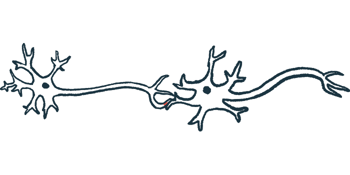
A feature of Parkinson’s is the formation and accumulation of toxic clumps of alpha-synuclein inside neurons. It’s been established that these clumps can “spread” in the brain, being transmitted between neighboring neurons. The gradual spread of this toxic protein throughout the central nervous system (CNS) — the brain and spinal cord — is thought to be a critical process driving the disease’ progression.
The currently accepted view for the damaging effects of the clumps and their spread is based on a “prion-like” mechanism — that is, if a little bit of toxic alpha-synuclein gets into a neuron, it can trigger the healthy protein inside the cell to form toxic clumps, which then drives cellular damage and death.
A research team led by scientists in Germany previously established that mice fed an insecticide called rotenone develop Parkinson’s-like disease. The animals show the spread of toxic alpha-synuclein aggregates through the CNS in a manner that’s comparable to how the disease progresses in people.
Testing ‘prion-like’ hypothesis of alpha-synuclein’s toxicity
To test the prion-like hypothesis, the researchers evaluated the effects of rotenone, paraquat (another Parkinson’s-associated insecticide), and toxic alpha-synuclein clumps in mice genetically engineered to not produce alpha-synuclein.
The team found that mice lacking alpha-synuclein performed significantly worse on motor function tests than their unaltered counterparts and their neurons showed abnormalities in mitochondria-derived energy production, which is consistent with the idea that some amount of alpha-synuclein protein is required for normal CNS function.
These mice didn’t develop Parkinson’s-like disease when exposed to rotenone, however. The animals showed similar numbers of dopamine-producing (dopaminergic) neurons — the nerve cells primarily affected in Parkinson’s — to healthy mice not exposed to the insecticide.
As such, “the presence of [alpha-synuclein] is necessary for PD [features] to progress,” the researchers wrote.
The researchers then took neurons from mice with and without alpha-synuclein and exposed them to toxic alpha-synuclein clumps in lab dishes. They observed similarly toxic effects on the dopaminergic neurons’ survival regardless of whether the nerve cells themselves already had any of the healthy protein inside them.
Similar findings were seen when the cells were treated with rotenone, suggesting “the presence of endogenous ASYN [the cell’s own alpha-synuclein] didn’t influence the toxicity” of alpha-synuclein clumps and rotenone, the team wrote.
The researchers also found that the toxicity of toxic alpha-synuclein on mitochondria was proportional to the amount of internalized protein. When toxic alpha-synuclein is administered to nerve cell fibers, it gets taken up and transported to the main nerve cell body, where it interacts with mitochondria and interferes with cellular energy production.
Analyses of survival rates of lab-grown neurons showed dopaminergic neurons were much more sensitive to toxic alpha-synuclein aggregates than other populations of nerve cells. Their sensitivity was similar regardless of whether they had their own healthy alpha-synuclein, however.
Similar results were obtained when the cells were treated with rotenone or paraquat.
These findings “challenge the current accepted hypothesis that [Parkinson’s] is a prion-like disease in which [internalized alpha-synuclein clumps] serve as seeds for the aggregation of the host’s endogenous [alpha-synuclein] and are the main drivers of [disease] progression,” the researchers wrote.
They emphasized that if the seeding effect of internalized alpha-synuclein clumps on nerve cells’ own alpha-synuclein protein was crucial to the disease-associated processes, “one would expect the lack of endogenous [alpha-synuclein] to reduce the toxicity of [alpha-synuclein clumps].”
While differences in the sensitivity to rotenone, paraquat, and alpha-synuclein clumps were detected between different types of neurons “we did not observe any differences in the sensitivity to these compounds or [alpha-synuclein]” between neurons with and without alpha-synuclein, the researchers wrote.
Effect on mitochondria increase oxidative stress
The results suggest the toxic effects of alpha-synuclein aggregates are driven by their effects on mitochondria, whereas prion-like spread “plays, if at all, a marginal role in this process,” the researchers wrote.
They hypothesized that alpha-synuclein’s effects on mitochondria cause dysfunction and increase a type of cellular damage called oxidative stress that promotes the aggregation of the healthy alpha-synuclein protein, which is then released by the neuron.
“Released [alpha-synuclein] re-starts the cycle when up-taken by [neighboring] neurons and triggers an inflammatory reaction,” the researchers wrote, adding that the results indicate that the “basis for the differences in the prevalence between PD-related non-motor symptoms and motor symptoms could be the difference in the susceptibility to external [damaging stimuli] (especially those affecting mitochondrial dysfunction and oxidative stress) between neuronal subtypes.”


Leave a comment
Fill in the required fields to post. Your email address will not be published.