‘Virtual Brain’ Could Lead to More Effective, Personalized DBS Use
Written by |
Using a personalized “virtual brain” could help in determining the optimal configuration of deep brain stimulation for people with Parkinson’s disease and other neurological disorders, a study suggests.
“We offer a computational model that holds the potential to be easily translated towards the individual patient level and used as a ‘sandbox’ model before future DBS [deep brain stimulation] surgeries,” its researchers wrote.
The study, “Virtual deep brain stimulation: Multiscale co-simulation of a spiking basal ganglia model and a whole-brain mean-field model with The Virtual Brain,” was published in Experimental Neurology.
Deep brain stimulation, or DBS, is a surgical treatment for Parkinson’s that involves implanting a device, called a neurostimulator, usually in the chest and leads with electrodes in specific brain areas for electrical stimulation. The overall aim is to help normalize electrical activity in the brain.

While it is well-established that DBS can help ease Parkinson’s motor symptoms, exactly how this intervention affects the brain’s electrical activity is incompletely understood. Its effects also likely vary from person to person depending on factors like where exactly in the brain a lead is placed.
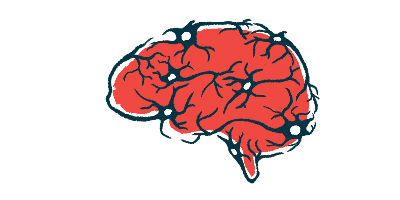
Scientists in Germany built upon previous research to devise a “virtual brain” that they could use to model DBS. Their model drew upon established knowledge about how different brain regions are connected, as well as the type of signals generated by DBS.
Notably, prior studies used similar methods in attempts to model certain brain regions. This model, in contrast, includes the entire brain.
“Compared to spiking models that encompass the BG [basal ganglia] regions only, our presented model can show whole-brain effects of stimulation going beyond the motor cortex,” the researchers wrote.
Instead of creating two models — a “Parkinson’s brain” and a “non-Parkinson’s brain” — the researchers also devised their model so it could be fit based on brain imaging data acquired in MRI scans. In this way, the virtual brain could be tailored to mimic the brain of a specific individual with Parkinson’s.
The team suggested that clinicians could “try out” different lead placements or DBS settings using this virtual model to find those most likely to benefit the patient before the start of surgery.
“Different strategies for DBS lead placements and configurations can be tested and evaluated,” the researchers wrote, adding, “the flexibility of the presented virtual model could help with finding the best therapy for each individual patient.”
They conducted a series of proof-of-concept tests with the model based on Parkinson’s brains undergoing DBS with different settings or locations. The scientists noted that, in all of these tests, DBS led to increased activity in the thalamus, which is located in the motor cortex region of the brain that, as its name implies, helps to control movement.
Prior research in patients also suggested that DBS increases activity in the thalamus, but the researchers stressed that differences in thalamus activity alone cannot explain all of Parkinson’s symptoms or all of the therapeutic effects of DBS.
Indeed, results from work with this model showed “whole-brain effects of stimulation going beyond the motor cortex,” the researchers reported.
“Future work needs to validate this model in larger patient cohorts and establish its link with clinical post-surgery improvement,” the team concluded.