Protein Clumps Marking Parkinson’s May Start in Digestive Tract
Early studies support link between damage to gut microbiome and disease

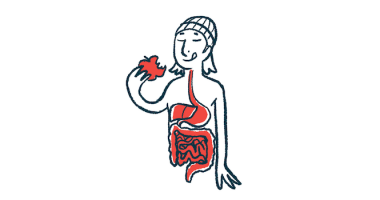
Toxic clumps of the alpha-synuclein protein, the underlying cause of neurodegeneration in Parkinson’s disease, may originate in the digestive tract before migrating to the brain, a study in mice reported, supporting previous work into a gut-brain axis in disease development.
Changes to microbes in the digestive tract, known as the gut microbiome, may facilitate alpha-synuclein clumping in the nerves that supply the gut, data also suggested.
The findings were in the poster “A new Parkinson’s disease model — Intracolonic rotenone causes alterations of gut microbiota and induces α-synuclein aggregation in the brain” presented at Neuroscience 2022, the Society for Neuroscience’s annual meeting, held Nov. 12–16 in San Diego.
This study was part of a group of investigations presented at the annual meeting examining the impact of the gut microbiome on neurological diseases like Parkinson’s, and on metabolic conditions such as obesity.
Damage to gut microbiome tied to alpha-synuclein clumps in gut, brain
“The neuroscience research presented today illustrates that, when it comes to metabolic and neurological disorders, we cannot target only the brain,” Sonia Villapol, PhD, an assistant professor of neurosurgery at Houston Methodist Research Institute, said in a press release from the Society for Neuroscience.
“Everything that happens in the gut has an impact on the brain,” Villapol said, adding that “a better understanding of interactions between the gut and the brain will bring great opportunities for the diagnosis, treatment, and prevention of diseases.”
In Parkinson’s, the progressive loss of neurons in the brain is linked to the misfolding and clumping of the alpha-synuclein protein. Such toxic aggregates promote damage to specialized neurons that produce the nerve-signaling molecule dopamine, also called dopaminergic neurons, in an area of the brain known as the substantia nigra. The loss of dopaminergic neurons triggers disease symptoms.
Increasing evidence supports changes in gut microbiota as a potential contributor to Parkinson’s. Patients show an imbalanced gut microbiota, and the fact that gastrointestinal problems can occur years before Parkinson’s onset suggest that the gut-regulating nervous system may be the first to be affected by the disease.
Notably, studies in mice suggest that alpha-synuclein clumps can form in the gut and spread to the brain, eventually causing Parkinson’s.
Since exposure to rotenone, a classic pesticide, has been linked to a higher the risk of Parkinson’s and shown to trigger Parkinson’s-like symptoms in animals, researchers at Rutgers Robert Wood Johnson Medical School, in New Jersey, looked at alpha-synuclein dynamics in mice after injecting rotenone into their colons.
Rotenone was administered into the colon’s initial parts (the proximal colon) for six weeks. About a half-year later, the team assessed the levels of alpha-synuclein aggregates in both the gut and the brain, as well as changes in dopaminergic neurons and motor function.
Compared with mice not given the pesticide (a control group), those exposed to rotenone showed increased alpha-synuclein production in the gut’s mucosal layer — the outer layer of digestive tract tissue that comes in contact with food and microbes. Staining of colon tissue confirmed the presence of alpha-synuclein aggregates in the neuron-containing layer directly below the mucosal layer.
Rotenone exposure also was linked to higher levels of a damaging form of alpha-synuclein called pS129, detected in the primary nerves that supply the digestive tract and in the brain’s substantia nigra. The number of dopaminergic neurons in this brain region was also significantly reduced in rotenone-exposed mice relative to control animals.
Problems in motor function were also evident in exposed mice, who took significantly less time to fall off a rotating rod.
Alpha-synuclein clumps may originate in the colon and propagate to the brain.
Molecular analysis demonstrated changes in the types of gut microbes that lasted more than 22 weeks (around five months) after rotenone administration.
The ratio of two bacteria types, Firmicutes and Bacteroidetes, was significantly higher in the rotenone group relative to the control group, indicating dysregulation of gut microbiota. A strong association was also seen between greater Lactobacillus abundance and fewer dopaminergic neurons.
These findings “indicate that abnormal [alpha-synuclein] may originate in the colon and propagate to the brain, leading to dopaminergic neuronal loss, and gut microbiota might contribute to this process,” the researchers wrote.
In another poster, titled “Low-Dose, Oral Insecticide Exposure Impairs Gastrointestinal Function and Disrupts Nigrostriatal Dopamine Circuitry in Mice,” researchers at Emory University, in Atlanta, provided further evidence of a gut-brain axis role in Parkinson’s.
In the study, a group of mice were orally exposed once a week to low-dose deltamethrin, an insecticide known to disrupt dopamine signaling.
Changes in dopamine pathways and altered motor behaviors were seen in the deltamethrin-exposed mice compared with a control mouse group. Deltamethrin exposure also triggered constipation-like symptoms in male animals, but not in females.
These results suggest that “exposure to deltamethrin in adulthood leads to altered dopamine handling in the gut and brain, rendering the system more vulnerable to [Parkinson’s-like features],” the researchers wrote.


