
Brain’s recycling system is blocked by mutation tied to Parkinson’s
Mutation found to aid in buildup of cellular debris in brain cells
Written by |

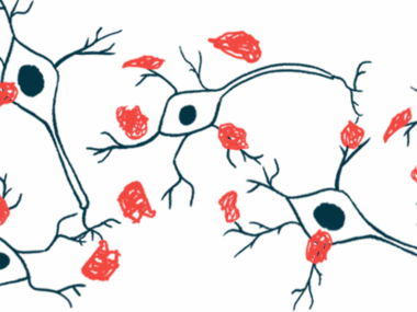
A mutation in the SH3GL2 gene that’s associated with an increased risk of Parkinson’s disease contributes to a buildup of cellular debris in brain cells, a study discovered.
According to researchers, that mutation was found to impair autophagy, an essential process by which cells recycle or breakdown components that are no longer needed or damaged. As a result, toxic debris build up in the brain and neurons die — two known hallmarks of Parkinson’s disease.
“Our team has found that a Parkinson’s disease-linked mutation in a gene called [SH3GL2] blocks the process by which the body and the brain recycle cell waste,” Adekunle Bademosi, the study’s first author and a postdoctoral researcher at the Queensland Brain Institute, in Australia, said in a university press release.
This discovery could change the way Parkinson’s is treated, according to Bademosi.
“It may be time to shift the treatment focus to autophagy as the mechanism underlying these disease hallmarks,” Bademosi said. “Exploring the use of compounds that induce or [suppress] autophagy could pave the way for new, more effective Parkinson’s drugs.”

Mutation in SH3GL2 gene linked to cellular debris in brain cells
The new study, “EndophilinA-dependent coupling between activity-induced calcium influx and synaptic autophagy is disrupted by a Parkinson-risk mutation,” was published in the journal Neuron.
Parkinson’s is marked by the progressive loss of neurons that produce the neurotransmitter dopamine, and in the early stages of the disease, impair synaptic function.
Neurotransmitters are chemical messengers used by neurons, or nerve cells, to communicate with each other. Synapses are the gaps where electric and molecular signals are transmitted from one neuron to the next.
In response to an electrical impulse, neurotransmitters are released from one neuron (pre-synaptic) into the synapse, crossing the gap to bind to receptors on the neighboring neuron (post-synaptic), stimulating another electrical signal.
Synapses are densely packed with proteins and other molecules that drive neurotransmitter release and signaling. Autophagy refers to the mechanism that eliminates used, unused, or damaged cell components, clearing cells of debris to maintain healthy function.
Disruptions in neuronal autophagy cause synaptic dysfunction and, ultimately, neurodegeneration.
Several proteins — including endophilinA1, known as EndoA1, which is encoded by the SH3GL2 gene — have been implicated in pre-synaptic autophagy. Many of these proteins are not involved in autophagy elsewhere in the nerve cell, research has shown.
EndoA1’s function is activated, in part, by an enzyme called LRRK2.
Variants in the gene that codes for LRRK2 have been associated with both familial and sporadic Parkinson’s. Moreover, a mutation in the SH3GL2 gene has been shown to increase Parkinson’s risk, as well as lead to increased activity of this gene.
However, the impact of these mutations on synaptic autophagy and Parkinson’s risk remains unclear.
“We knew we could induce autophagy in cells by starving them of amino acids [proteins’ building blocks] and the subsequent breakdown of debris tells a protein called EndoA to approach the cell membrane and begin the recycling process,” Bademosi said. EndoA is the fruit fly version of EndoA1.
Using the fruit fly model, the researchers found that synaptic autophagy also was independently stimulated by an electrical signal, which triggered the influx of positively charged calcium ions and activated EndoA.
“Now we’ve also seen that regular signals between neurons in the brain starts EndoA-induced autophagy when the electric impulses trigger the release of proteins or neurotransmitters at synapses,” Bademosi said.
Calcium-mediated EndoA activation was then shown to rely upon certain negatively charged amino acids, located in a flexible, unstructured part of the protein.
The team found that calcium influx stimulated EndoA to move from the edges of the cell into the vicinity of the pre-synapse membrane. Mutating these amino acids to remove their negative charge made EndoA less flexible and eliminated its response to calcium influx.
The ultimate result was impaired synaptic autophagy that led to neurodegeneration, including damage to dopamine-producing neurons.
Our team has found that a Parkinson’s disease-linked mutation in a gene called [SH3GL2] blocks the process by which the body and the brain recycle cell waste.
Data also showed that EndoA-induced synaptic autophagy was independently promoted by either calcium influx or LRRK2-dependent activation of EndoA. These “are independent, parallel events that can override each other,” the team wrote.
The researchers then demonstrated that the Parkinson’s-associated SH3GL2 mutation, called G276V, impaired calcium-mediated synaptic autophagy in the fruit fly model and in lab-grown human dopamine-producing neurons.
“Unfortunately, when the [SH3GL2] gene is affected in Parkinson’s, the protein EndoA becomes insensitive to [electrical signals] at the synapse, and the debris that should be thrown out for recycling builds up instead,” Bademosi said.
While it is still unknown how impaired synaptic autophagy leads to neuron loss, “our work does show a correlation between defective (too much and too little) synaptic autophagy and neurodegeneration, also of dopaminergic neurons,” the team wrote.
“This work reveals that EndoA links neuronal activity-dependent [calcium] influx to synaptic autophagy and that a human mutation in the unstructured loop region of EndoA conferring risk to [Parkinson’s Disease] disrupts this process,” they concluded.
Leave a comment
Fill in the required fields to post. Your email address will not be published.