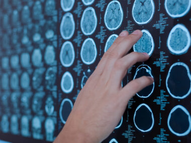
Brain imaging method may aid mild traumatic brain injury diagnosis
mTBIs are associated with 50% higher risk of developing Parkinson's
Written by |

A new brain imaging method may help diagnose mild traumatic brain injury (mTBI), which according to some studies can be associated with a 50% higher risk of developing Parkinson’s.
Available methods, like magnetic resonance imaging (MRI), leave most cases of mTBI, or concussions, undiagnosed. They occur when a physical injury such as a violent blow or jolt to the head leads to brain damage.
“70-90% of reported TBI cases are categorized as ‘mild,’ yet as many as 90% of mTBI cases go undiagnosed, even though their effects can last for years and they are known to increase the risk of a host of neurological disorders including … Parkinson’s disease,” Samir Mitragotri, PhD, study’s senior author in whose lab the research was performed, said in a press release. The method is described in “Preclinical characterization of macrophage-adhering gadolinium micropatches for MRI contrast after traumatic brain injury in pigs,” in Science Translational Medicine.
mTBI-induced brain inflammation produces signals that attract macrophages, a type of immune cell that infiltrates the brain in response to inflammation. Most people with a mTBI don’t get a proper diagnosis, however, which can exacerbate the injuries and lead to further damage.
An undiagnosed mTBI may lead to “invisible” brain inflammation that increases the injury’s effects, which raise the risk of developing several neurological conditions, including Parkinson’s and chronic traumatic encephalopathy, a neurodegenerative condition that affects more than 90% of American pro football players. The researchers at Mitragotri’s lab, in the Wyss Institute at Harvard University, relied on this understanding and previous experience with immune cells to reach a better diagnosis method for mTBI.
Improving imaging in the brain
“Our previous projects have focused on controlling the behavior of immune cells or using them to deliver drugs to a specific tissue,” said Lily Li-Wen Wang, PhD, the first author of the study. “We wanted to exploit another innate ability of immune cells — homing to sites of inflammation in the body — to carry imaging agents into the brain, where they can provide a visible detection signal for mTBI.”
The technique involves attaching gadolinium, a standard MRI contrast agent, to macrophages via hydrogel-based microparticles. For gadolinium to be used as a contrast agent, it must interact with water and the researchers’ previous macrophage-attaching technology used a molecule that repels water. For this reason, they had to develop new hydrogel microparticles, which they called M-GLAMs, that can produce a strong gadolinium-mediated MRI signal and form a stable attachment to both mouse and pig macrophages.
Teaming up with researchers and clinicians at Boston Children’s Hospital, they showed that injecting mouse M-GLAMs macrophages in mice didn’t lead to them accumulating in the kidneys, but they remained in the body for more than 24 hours without adverse side effects. Gadavist, an existing gadolinium-based contrast agent, does accumulate in the kidneys, which can cause health risks for patients with kidney disease.
Moreover, in a pig model of mTBI, pigs that received M-GLAMs displayed a significant increase in the intensity of gadolinium in the choroid plexus, a major conduit of immune cells in the brain, while those injected with Gadavist didn’t, despite increased inflammation-related macrophage density in the brains of both groups of animals.
“Another important aspect of our M-GLAMs is that we [can] achieve better imaging at a much lower dose of gadolinium than current contrast agents- 500-1,000-fold lower [than] Gadavist,” said Wang. “This could allow the use of MRI for patients who are currently unable to tolerate existing contrast agents, including those who have existing kidney problems.”
Although the method can’t identify the exact location of the brain injury or inflammation, it may offer a faster and more effective way to detect inflammation in mTBI patients.
“Our results suggest that macrophage-adhering GLAMs could facilitate mTBI diagnosis,” the researchers wrote.
Moreover, if coupled with a new treatment modality developed by the same team that uses macrophages attached to microparticles with anti-inflammatory molecules, it’s possible to minimize mTBI-related damage and accelerate recovery.
The researchers submitted a patent application for their technology and hope to bring it to market shortly. They are also exploring collaborations with biotech and pharmaceutical companies to accelerate its development into clinical trials.
“This work demonstrates just how much potential is waiting to be unlocked within the human body for a variety of functions: monitoring health, diagnosing problems, treating diseases, and preventing their recurrence,” said Donald Ingber, MD, PhD, Wyss Founding’s director.


