Toxic alpha-synuclein protein seen to alter energy use in nerve cells
New technique shows diseased nerve cells burn glucose faster than healthy ones
Written by |

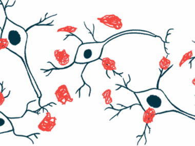
Toxic clumps of the protein alpha-synuclein change the way the brain cells most affected by Parkinson’s disease utilize energy, a study shows.
Its researchers wanted a clearer understanding of how “key metabolic processes” are affected by the protein’s aggregation, damaging “vulnerable” nerve cells within the brain, they wrote. To do so, they developed a technique to peer inside brain cells.
“Although high metabolic needs are considered as an important factor in the degeneration of specific neuronal populations, this aspect has rarely been addressed,” the scientists wrote.
Their study, “Stable isotope labeling and ultra-high-resolution NanoSIMS imaging reveal alpha-synuclein-induced changes in neuronal metabolism in vivo,” was published in Acta Neuropathologica Communications.
Study into how protein clumps might open dopaminergic neurons to damage
Parkinson’s is caused by the progressive dysfunction and death of dopamine-making nerve cells, which are mainly located in a part of the brain called the substantia nigra.
It’s not known exactly what causes these nerve cells to sicken and die, but clumps of alpha-synuclein are thought to play a major role. Alpha-synuclein aggregates are found in the brains of virtually all with Parkinson’s disease, and these clumps are known to be toxic to nerve cells.
Exactly how alpha-synuclein clumps damage nerves — and why this damage seems to hit dopamine-making cells in the substantia nigra harder than other brain cells — is not fully understood.
To investigate, researchers in Switzerland used a rat model where alpha-synuclein clumps were induced on one side of the brain. The other side — the left hemisphere — remained healthy for comparison.
Rats then were fed water containing radioactively labeled molecules of glucose, the sugar molecule that cells burn in order to generate energy. The scientists used an advanced imaging technique called NanoSIMS to track the uptake of the radioactive glucose by individual brain cells.
By combining the NanoSIMS images with detailed pictures of the cell taken by an electron microscope, the researchers could use this technique to assess the movement of sugar molecules within individual structures inside the cells.
“This study shows the potential of the NanoSIMS technology to reveal metabolic changes in the brain, with unprecedented resolution, at the subcellular level. It hands us a tool to study early pathological changes occurring in vulnerable neurons as a consequence of alpha-synuclein accumulation, a mechanism directly linked to Parkinson’s disease,” Bernard Schneider, PhD, a study co-author at Ecole Polytechnique Fédérale de Lausanne, said in a university press release.
In initial tests, the team noticed that dopamine-making neurons in the substantia nigra tended to take up more sugar than other brain cells. This finding adds to evidence indicating that these nerve cells usually require a lot of energy to function, which may help to explain why they are more vulnerable with Parkinson’s, the researchers said.
The initial uptake of sugar was comparable in dopamine-making neurons with or without alpha-synuclein clumps, results showed. However, cells with toxic protein clumps tended to burn through the sugar a lot faster than their healthy counterparts, suggesting a higher metabolic activity overall in the diseased neurons.
Alpha-synuclein aggregates also caused changes at the subcellular level. In particular, the researchers found a lower uptake of the sugar molecule in mitochondria — cellular structures needed to generate cell energy — as well as the Golgi apparatus, a cellular structure used to help make new cell components.
These findings imply that dysfunction in the mitochondria and Golgi may be major reasons for the toxic effects of alpha-synuclein within nerve cells, the researchers said.
More broadly, their approach could be used to investigate metabolic problems in other neurological diseases, they noted.
“This powerful approach can be used to reveal changes in the metabolic activity of neuronal subpopulations at the subcellular level and compare normal and disease conditions,” the scientists wrote, adding that it “can help interrogate the dynamics of neuronal metabolism in the normal brain and investigate the metabolic changes underlying neurodegenerative diseases.”


