Better Diagnostic Imaging for Parkinson’s Goal of Australian Research Team
Written by |

A high-resolution imaging approach that could help in diagnosing Parkinson’s disease by enabling physicians to more easily visualize the substantia nigra, the brain region responsible for controlling movement and whose nerve cells are gradually lost to this disease, is the goal of a research group in Australia.
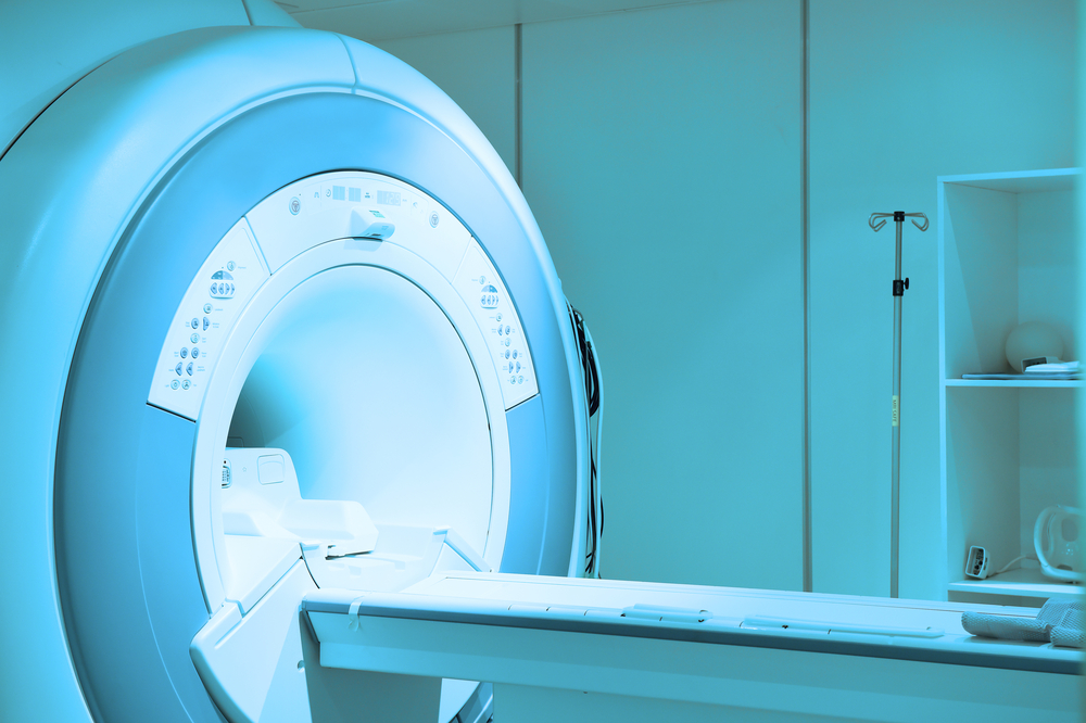
The method, which aims to builds on advances in magnetic resonance imaging (MRI), is being developed by Gabrielle Todd, PhD, and Shayne Chau, neuroscience and medical imaging specialists at the University of South Australia (UniSA), in collaboration with physicians from the Flinders Medical Centre in metropolitan Adelaide.
According to Todd, current positron emission tomography (PET) and single-photon emission computed tomography (SPECT) imaging ably allow clinicians to evaluate the degree of neurodegeneration in patients’ brains.
However, both methods require patients to be injected with radioactive substances, known as radiotracers, to improve contrast and image quality.
“The significant challenges associated with manufacturing radiotracers and the high cost severely limits patient access to this technology in Australia and elsewhere,” Todd said in a university press release. “Due to the lack of an accurate and accessible diagnostic test for Parkinson’s disease, this results in a high rate of misdiagnosis.”
MRI is relatively safe and much more accessible. But because it does not use radiotracers, only very experienced radiologists can tell the difference between a normal and abnormal substantia nigra based on a brain MRI scan, she said. Again, this risks a misdiagnosis.
With her team, Todd is working to develop and test an accurate but easy-to-use imaging approach, one with the advantages of conventional MRI. At the same time, it would allow physicians to objectively measure the substantia nigra of a patient, and compare data with that from a reference control population.
“Radiologists rarely see what a normal substantia nigra looks like in healthy people and we don’t currently know if the appearance of the substantia nigra changes with age or whether it differs between males and females,” she said. “Collectively, we hope to make it easier for radiologists to learn about and interpret abnormal substantia nigra MRI findings and to provide neurologists with more certainty about the patient diagnosis.”
Newly diagnosed Parkinson’s patients and healthy adults will be recruited to undergo a brain MRI scan, as well as movement, memory, and cognitive function tests, to establish objective guidance.
Marty Edwards, a prominent winemaker diagnosed with Parkinson’s at 41, is donating proceeds of his new wine label — Silver Lining Wines — to help fund the work.
“This project will increase patient access to cutting edge medical imaging in both metropolitan and regional communities throughout Australia and other countries. It will enable faster and more accurate diagnosis of Parkinson’s disease and so allow people to be treated earlier to help control their symptoms,” Edwards said.
“Being associated with a project that can provide hope, and arm clinicians with more tools to help patients, is something that I’m passionate about. Hopefully we’ll create a ‘silver lining’ for others that are starting their journey with Parkinson’s disease,” he added.


