Tracers help ID reactive astrocytes in Parkinson’s brain
Imaging markers show sites of inflammation, study finds
Written by |
Two imaging markers can help pinpoint sites of brain inflammation in people with Parkinson’s disease, according to a study.
The findings shed light on the role of brain inflammation in disease progression and can help pave the way for more precise diagnostics and targeted therapies.
“We are excited about the potential of these imaging tracers to advance our understanding of Parkinson’s disease,” Amit Kumar, PhD, research specialist at Karolinska Institutet in Stockholm, said in an institute news story. “Our goal is to develop strategies that can help manage and treat this condition more effectively.”
The study, “Mapping reactive astrogliosis in Parkinson’s brain with astroglial tracers BU99008 and Deprenyl: New insights from a multi-marker postmortem study,” was published in Alzheimer’s & Dementia.
Parkinson’s disease is caused by the death and dysfunction of nerve cells in the brain that are responsible for the production of dopamine, a signaling molecule that plays a role in motor control. But evidence points to the dysfunction of other cells, namely astrocytes, as key contributors to Parkinson’s progression by promoting brain inflammation, or neuroinflammation.

Reactive astrogliosis
The researchers looked at reactive astrogliosis, a process in which astrocytes — star-shaped cells found in the brain and spinal cord — undergo structural and functional changes in response to injury, disease, or inflammation. Reactive astrogliosis is a hallmark of several neurodegenerative conditions, including Alzheimer’s disease and multiple sclerosis. Astrocytes play a supportive role for neurons in processes such as regulating the levels of brain chemical messengers, repairing brain tissue, and providing structural support.
While evidence from postmortem brain tissue samples from Parkinson’s patients supports a role for reactive astrogliosis, the lack of specific biomarkers for this process has limited its characterization.
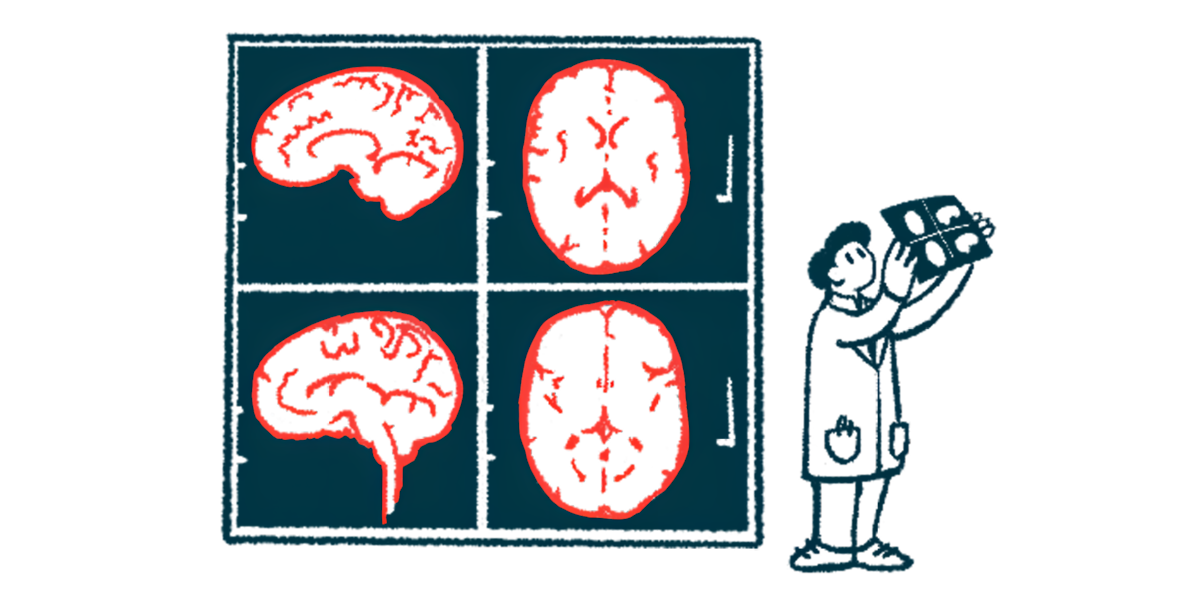
Two imaging biomarkers, BU99008 and Deprenyl, were recently developed as markers of reactive astrocytes to be used during positron emission tomography scans. This type of imaging uses a radioactive molecule, called a tracer, that is injected into the body and binds to specific proteins to make them visible and show areas of disease.
“Despite their immense potential these tracers have not been thoroughly studied and validated in [Parkinson’s disease],” the researchers wrote.
The researchers used both tracers in postmortem brain tissue of patients with Parkinson’s, comparing it to tissue from people without the disease.
They analyzed four brain regions — frontal cortex, temporal cortex, caudate nucleus and putamen — in postmortem tissues of four Parkinson’s patients and four people without the disease, who served as controls.
While these brain areas have their unique functions, all are involved in various aspects of cognitive processing and behavior.
The analysis showed distinct binding patterns in Parkinson’s patient’s brains compared with controls, indicating various types of binding sites with unique affinities and ratios in different regions of the brain.
Both BU99008 and Deprenyl were able to detect reactive astrogliosis, suggesting that these tracers could potentially serve as indicators of reactive astrogliosis in Parkinson’s patients’ brains.
According to the researchers, “the tracers are binding to different subpopulations or specific state of astrocytes, as we also suggested in our previous studies with [Alzheimer’s disease] brains.”
The presence of astrogliosis was further evidenced by the presence of astrocytes that were positive for two specific proteins, GFAP and ICAM-1, in the brains of those with Parkinson’s. These molecules are used as markers of advanced/end stages of Parkinson’s, and are involved in the activation of immune responses as a consequence of inflammation.
“These findings are crucial as they provide a better understanding of the role of reactive astrogliosis in Parkinson’s disease,” Kumar said. “By using these novel tracers, we can now study these changes in greater detail, which could lead to new diagnostic interventions and treatments.”
The researchers plan to further explore these and other novel inflammatory markers and their roles in Parkinson’s progression.