Proteins pair to help alpha-synuclein spread in brain: Study
Aplp1, Lag3 allow toxic aggregates into nerve cells
Written by |
Two proteins, Aplp1 and Lag3, pair to allow toxic aggregates of alpha-synuclein — a hallmark of Parkinson’s disease — into nerve cells and help it spread across the brain, according to a study in mice.
“Now that we know how Aplp1 and Lag3 interact, we have a new way of understanding how alpha-synuclein contributes to the disease progression of Parkinson’s disease,” Xiaobo Mao, PhD, associate professor of neurology at Johns Hopkins Medicine and the study’s first author, said in a Hopkins press release.
The study, “Aplp1 interacts with Lag3 to facilitate transmission of pathologic [alpha]-synuclein,” was published in Nature Communications.
The team also found that using an antibody against Lag3 blocked the protein’s interaction with Aplp1, keeping alpha-synuclein from being taken into nerve cells and causing neurodegeneration.
The findings suggest that “targeting this interaction with drugs could significantly slow the progression of Parkinson’s disease and other neurodegenerative diseases,” Mao said.

Alpha-synuclein misfolds in Parkinson’s
Lag3 is already a target of a therapy, Opdualag (relatlimab/nivolumab), approved in the U.S. and Europe to treat a type of skin cancer.
In Parkinson’s disease, the protein alpha-synuclein misfolds and aggregates into small structures known as fibrils. These fibrils coalesce to form Lewy bodies, which are a hallmark of the disease. Lewy bodies are found in the brains of people with Parkinson’s, where they gradually drive neurodegeneration.
Once the first fibrils are formed, they can spread from one neuron, or nerve cell, to another across the gut and into the brain, triggering more neurons to form Lewy bodies. This is believed to be a key driver of Parkinson’s and how fast it progresses over time.
Earlier work by Mao and other researchers showed that alpha-synuclein fibrils interlock with Lag3, a protein known for its role in the control of the immune response, and hijack their way into neurons. Now, Mao’s team discovered that alpha-synuclein needs yet another protein to spread around.
“Our work previously demonstrated that Lag3 wasn’t the only cell surface protein that helped neurons absorb alpha-synuclein, so we turned to Aplp1 in our most recent experiments,” said Valina Dawson, PhD, neurology professor and co-director of the neuroregeneration and stem cell programs at Johns Hopkins Institute for Cell Engineering.
Aplp1 is a beta-amyloid precursor-like protein known to bind to alpha-synuclein. Like alpha-synuclein, beta-amyloid forms toxic clumps in neurons, causing their death in Alzheimer’s, another neurodegenerative disease. However, the role of Aplp1 in the spread of alpha-synuclein was unknown.
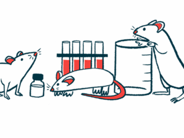
The researchers turned to a mouse model in which the gene coding for Aplp1 had been turned off, or knocked out. That meant that unlike wild-type (healthy) mice, knock-out mice were unable to produce Aplp1.
To test if Aplp1 helps in alpha-synuclein uptake, the researchers labeled preformed fibrils with a dye that lights up when it gets inside cells. They injected the labeled fibrils into the mice’s brains, and then tracked them using a microscope.

Implications for Parkinson’s research
Compared with wild-type neurons, those of knock-out mice took up about half as many alpha-synuclein fibrils into endosomes, sac-like structures that engulf proteins and other materials into cells. However, adding Aplp1 back increased alpha-synuclein uptake.
To find out if alpha-synuclein needs Aplp1 to cause neurodegeneration, the researchers measured the amount of pS129, an abnormal version of alpha-synuclein that folds into a shape that tends to form toxic clumps. They found that the levels of pS129 were reduced by about 60% in knock-out neurons.
Similar observations were made in mice that lacked both Aplp1 and Lag3. In these mice, pS129 levels were reduced by about 90% compared with wild-type mice. At the same time, dopaminergic neurons — those responsible for producing dopamine, a chemical involved in motor control — were largely preserved.
As a result, mice lacking both Aplp1 and Lag3 were faster than wild-type mice at crawling down a pole. When placed in a hollow glass cylinder to check for spontaneous movements, they also moved more freely than wild-type mice.
Weekly injections of 410C9, an antibody against Lag3 that was found to block its interaction with Aplp1, reduced the number of alpha-synuclein fibrils taken up by neurons and decreased their spread, preventing the death of dopaminergic neurons.
The antibody, which was found to be able to reach the brain, also eased motor symptoms of Parkinson’s. Besides crawling faster down a pole, mice treated with 410C9 also performed better in a grip strength test than those given a placebo.
“The anti-Lag3 antibody was successful in preventing further spread of alpha-synuclein seeds in the mouse models and exhibited better efficacy than Lag3-depletion because of Aplp1’s close association with Lag3,” said Ted Dawson, MD, PhD, who co-led the study.
The study “provides additional targets for therapeutic strategies aimed at preventing neurodegeneration in Parkinson’s,” the researchers wrote.
“While our study mainly focused on the neuronal function of Lag3-Aplp1, it will be important to determine if these proteins also work closely in other cell types,” they added.