
PET radiotracer captures inflammation in human brain
First-in-human study uses marker that binds to COX-2 enzyme
Written by |
A new positron emission tomography (PET) radiotracer can accurately visualize inflammation within the healthy human brain, a first-in-human study found.
By using a marker that binds to COX-2, a key enzyme associated with brain inflammation or neuroinflammation, researchers were able to quantify low levels of this enzyme in healthy individuals.
“Neuroinflammation plays a critical role in many neurological and psychiatric diseases, including Alzheimer’s disease, Parkinson’s disease, and major depressive disorder,” Robert B. Innis, MD, PhD, senior investigator in the molecular imaging branch of the National Institute of Mental Health at the National Institutes of Health, said in a Society of Nuclear Medicine & Molecular Imaging press release. “This could be a game-changer for personalized medicine and therapeutic development.”
The study, “PET Quantification in Healthy Humans of Cyclooxygenase-2, a Potential Biomarker of Neuroinflammation,” appeared in The Journal of Nuclear Medicine, which is published by the society.
The ability to image neuroinflammation in a living human brain has sparked interest among neuroscientists, as it enables earlier disease detection, monitoring of disease progression, and assessment of anti-inflammatory treatments in neurological disorders.

COX-2 enzyme and neuroinflammation
One potential biomarker for brain inflammation is COX-2, an enzyme that’s found in small amounts in nerve cells and that rapidly increases in response to inflammation. While its precise functions in the brain remain unknown, its density may serve as a measurable indicator of neuroinflammation “even if it is not a mediator of the inflammatory process” itself, the researchers wrote.
“While COX-2 has been widely studied in peripheral inflammation, its role in neuroinflammation has been difficult to quantify in vivo,” Innis said. “We sought to establish a non-invasive imaging method to measure COX-2 in the living brain to enable earlier disease detection, monitor disease progression, and assess anti-inflammatory treatments.”
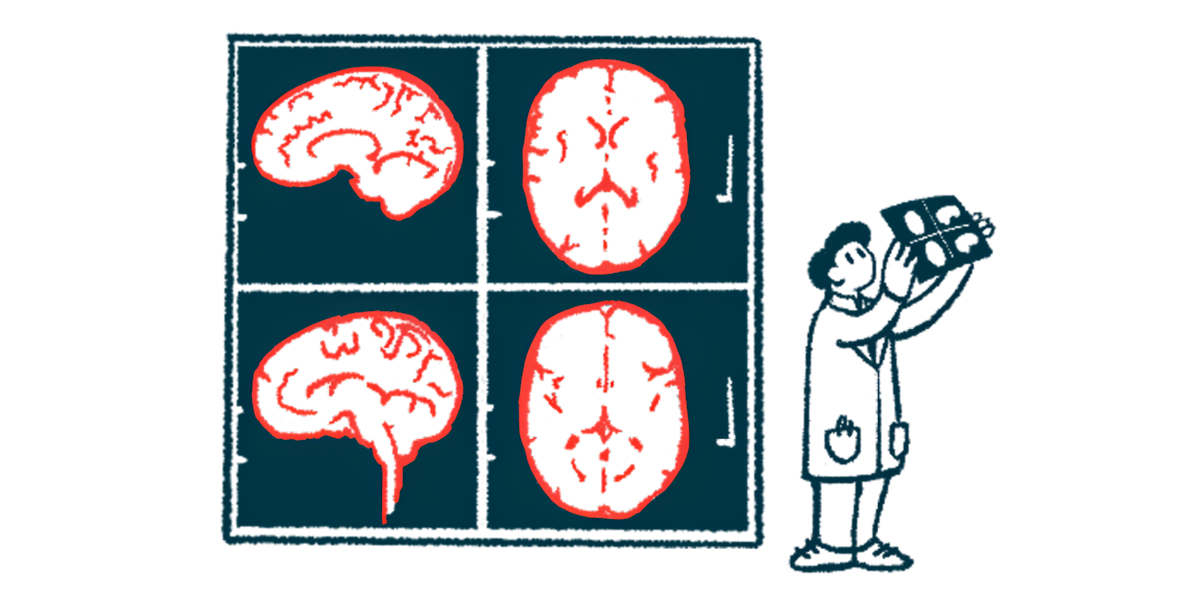
To achieve this, the researchers developed a new PET tracer called 11C-MC1. A PET tracer is a radioactive substance that helps create images of how the body works during a PET scan.
Once administered, 11C-MC1 travels through the bloodstream and crosses the blood-brain barrier, a protective membrane that keeps harmful substances from the brain, binding to COX-2. When 11C-MC1 binds to COX-2, its radioactive tag emits a signal that a PET scan captures to create a detailed image of COX-2 distribution in the brain.
Before testing its ability to detect COX-2 in healthy human brains, 11C-MC1 was tested in animal models to ensure it specifically bound to COX-2 and not to its closely related enzyme, COX-1.
Researchers used genetically modified mice expressing the human COX-2 gene. After injecting them with ¹¹C-MC1, PET scans showed a strong signal, confirming the tracer’s binding to COX-2. When the mice received either the tracer without its radioactive tag (MC1) or the COX-2 inhibitor celecoxib, brain uptake of ¹¹C-MC1 dropped by 92% and 74%, respectively, demonstrating its specificity. In contrast, the COX-1 inhibitor PS13 had no effect, confirming that the tracer does not bind to COX-1.
Subsequently, 27 healthy human volunteers were injected intravenously (into the vein) with 11C-MC1 and imaged with PET scans. The tracer efficiently crossed the blood-brain barrier, rapidly entering the brain and peaking after four minutes.
To confirm the specificity of 11C-MC1 for COX-2, 10 participants each received a PET scan at the beginning of the study followed by a second scan after taking celecoxib, “a preferential COX-2 inhibitor,” the researchers wrote. The observed reduction in 11C-MC1 binding demonstrated that the tracer selectively targets COX-2. To assess the reliability of 11C-MC1 in quantifying COX-2 levels, 17 participants underwent PET scans twice.
“This study found that the PET radioligand [11C-MC1] had adequate sensitivity to quantify COX-2 expression in the healthy human brain, making it a promising biomarker of neuroinflammation,” the researchers wrote. “These findings strongly support the notion that [11C-MC1] has adequate sensitivity to detect elevated COX-2 levels that may occur in various brain disorders.”
This noninvasive method opens the door for the use of COX-2 PET imaging in both clinical and research settings, potentially enhancing diagnostic precision and treatment strategies for neuroinflammatory conditions, including Alzheimer’s and Parkinson’s. “ It also demonstrates the potential for developing other PET tracers to investigate neuroinflammation, broadening the applications of nuclear medicine in neurology and psychiatry,” Innis said.
Leave a comment
Fill in the required fields to post. Your email address will not be published.