
Cell model recreates dynamics of alpha-synuclein clumping: Study
Model aims to mimic distinct ways protein aggregates form, spread in living brain
Written by |
A new cell model of Parkinson’s disease is better able than others at capturing the complexity of the toxic protein clumps that characterize the disorder and to do so with useful speed, a study reports.
“This technology will pave the way for rapidly developing ‘personalized stem cell models’ from individual patients,” Isabel Lam, PhD, the study’s co-first author and a research fellow in neurology at Brigham and Women’s Hospital in Boston, said in a news story from Harvard University.
These models “are already being used to efficiently test new diagnostic and treatment strategies ‘in a dish’ before jumping into clinical trials so we target the right drug to the right patient,” Lam added.
The study, “Rapid iPSC inclusionopathy models shed light on formation, consequence, and molecular subtype of [alpha]-synuclein inclusions,” was published in Neuron.

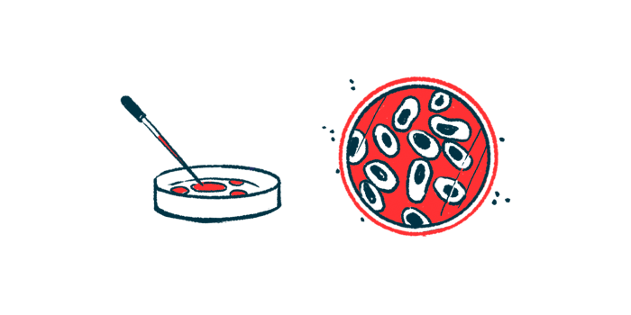
Alpha-synuclein clumps can take many forms within a brain and its cells
Parkinson’s is characterized by abnormal clumps of the protein alpha-synuclein in brain cells, which are toxic and thought to play a central role in initiating and driving disease processes. Clumps of alpha-synuclein also can take many forms in different parts of the brain, even within the same cell. The consequences of these different types of clumps are poorly understood.
“The problem is that the way protein clusters form in PD [Parkinson’s disease] looks different in different patients, and even in different brain cells of the same patient,” said Vikram Khurana, MD, PhD, chief of the hospital’s movement disorders division and the study’s principal investigator. “This begs the question: How do we model this complexity in the dish? And how do we do it fast enough for it to be practical for diagnostics and drug discovery?”
Scientists have created Parkinson’s models of nerve cells generated from patient-derived stem cells, then grown in a dish and studied. While these models provide some useful insights, their major drawback is in not accurately capturing the dynamics of alpha-synuclein clumping that are seen in the human brain.
An international research team, led by scientists at the Boston hospital, developed a new model to better capture these protein aggregation dynamics. Put simply, the model works by inducing stem cells to grow into brain cells while also engineering the cells to promote alpha-synuclein clumping.
“We sought to assess how quickly we could make human brain cells in the lab that give us a window into key processes occurring in the brains of patients with Parkinson’s disease and related disorders,” Khurana said. “And, unlike previous models, we wanted to do this in a short enough timeframe for these models to be useful for high-throughput genetic and drug screens and easy enough for many labs to use across academia and industry.”
Model triggers clumping in two ways, with aggregates showing distinct features
The protein clumping could be triggered either by genetically engineering cells to express clumping-prone protein within the cell, or by treating the cell with preformed protein clumps — mimicking two distinct ways these clumps can form and spread within the brain.
A notable takeaway from these experiments was that clumps formed in these different ways showed distinct molecular features, such as different types of other proteins and fatty molecules within the aggregates.
Findings suggest that viewing disorders like Parkinson’s as conditions associated with one type of protein clumping is too simplistic, the researchers said. Instead, these diseases “should be viewed as ‘polyproteinopathies’ [disorders of multiple proteins] in which the misfolding of one protein like [alpha-synuclein] leads to a multitude of misfolding, redistribution, and protein-sequestration events,” they wrote.
Researchers noted that, while their initial experiments focused mainly on alpha-synuclein, this model can be adapted to study other proteins like amyloid-beta and tau, which characteristically form clumps in the brains of people with Alzheimer’s disease.
The model currently is being used to help advance research into Parkinson’s, the scientists noted.
“In one key application, we are utilizing this technology to identify candidate radiotracer molecules to help us visualize alpha-synuclein aggregation pathologies in the brains of living patients we see in the clinic,” said Alain Ndayisaba, MD, the study’s other first author and a research fellow in neurology at Brigham and Women’s Hospital.


Leave a comment
Fill in the required fields to post. Your email address will not be published.