Alpha-synuclein lifts up at cell membranes; may promote clumping
When protein is at low levels, it remains flat against membrane surface
Written by |

When the alpha-synuclein protein is present at high concentrations around cellular membranes, it lifts up rather than lying flat against the membrane, making it more prone to clumping, a study shows.
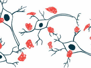
The finding may offer insight into the molecular mechanisms that drive Parkinson’s disease, where toxic clumps of alpha-synuclein accumulate in the brain.
The methods used in this study to follow alpha-synuclein motions in a model of a nerve cell’s surface may help “screen for effective therapies to prevent the disease,” Steven Roeters, PhD, one of the study’s authors at Aarhus University, Denmark, said in a press release. Roeters is a research associate at Amsterdam UMC, in the Netherlands. The study, “Elevated concentrations cause upright alpha-synuclein conformation at lipid interfaces,” was published in Nature Communications.
The causes of Parkinson’s aren’t fully understood, but alpha-synuclein clumps in the brain are thought to play a central role. These aggregates can be toxic to nerve cells, but the molecular details about how these clumps form remain obscure.
All human cells are surrounded by a membrane made of fatty molecules, or lipids. Being near a lipid membrane can prompt alpha-synuclein proteins to clump.
“The relevance of the aggregation [facilitation] by lipids is substantiated by the presence of lipids in Lewy bodies,” the researchers wrote. Lewy bodies are complex fiber-like, alpha-synuclein aggregates that are a hallmark of Parkinson’s.
What promotes alpha-synuclein protein clumping?
Figuring out what prompts this lipid-induced clumping was the aim of Roeters and his colleagues at Aarhus University, where they used a technique called vibrational sum-frequency generation (VSFG) to examine these molecular events in greater detail. In VSFG, two lasers are shone at different frequencies at the interface between a membrane and a protein sample, then how the lasers reflect off the sample are measured. Advanced computer algorithms can interpret the data to draw conclusions about the alpha-synuclein’s movement.
“We have developed an effective approach for extracting structural information from experimental VSFG spectra … [and] we [applied] it to investigate [alpha-synuclein] binding to lipid surfaces, from low concentrations — which have been studied previously using other methods — to physiological and elevated protein concentrations,” the researchers wrote.
They found that when alpha-synuclein proteins are present at low levels, like what’s typically seen in the body, they lie flat against the membrane surface. In this position, individual proteins don’t touch each other much, so they aren’t likely to clump.
But when there are high concentrations of alpha-synuclein, the proteins start to lift up away from the membrane. This causes more alpha-synuclein proteins to touch each other, which can set the stage for clumping.
“The upright orientation … provides an opportunity for [alpha-synuclein] to engage in more extensive intermolecular contacts and may pave the way for subsequent aggregation,” the researchers wrote. “This molecular mechanism can explain both the concentration-dependent aggregation [facilitation] by lipids and the aggregation-related toxicity observed at elevated [alpha-synuclein] concentrations.”
The findings and method can be used to closely follow the dynamics of alpha-synuclein misfolding and orientation in the presence of potential treatments, which may help accelerate drug development for Parkinson’s, noted the scientists, who plan to extend this research by testing how alpha-synuclein behaves in response to other surfaces and materials.
“We plan to examine the interactions of [alpha-synuclein] with artificial materials, such as microplastics to understand the impact of environmental conditions on Parkinson’s,” said Tobias Weidner, PhD, the study’s senior author and professor at Aarhus University.


