
Scientists Discover Mitochondria Recycling Method Likely Involved in Parkinson’s
Written by |
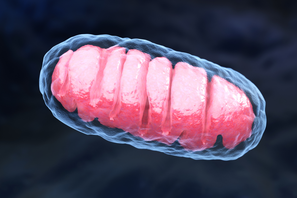
Tatiana Shepeleva/Shutterstock
An alternative method of recycling mitochondria — the cell’s powerhouses — that involves pushing damaged mitochondria out of cells, seems to play a role in Parkinson’s disease, according to a recent study.
Researchers have found evidence in cellular and animal models of disease that this process is enhanced in the absence of parkin, a protein that — when dysfunctional — is known to trigger the onset of hereditary forms of Parkinson’s.
Likewise, when researchers analyzed body fluid samples from patients carrying mutations that compromised parkin’s normal function, they discovered this alternative method of mitochondria recycling also was increased in those patients.
“These are striking results that demonstrate a novel mechanism for clearing defective mitochondria and an important role for mitochondrial homeostasis in the onset of Parkinson’s disease. These findings may lead to the development of new biomarkers and therapeutics,” Hideki Mochizuki, MD, PhD, professor at Osaka University in Japan, and corresponding author of the study, said in a press release.
The findings were reported in the study, “Alternative mitochondrial quality control mediated by extracellular release,” published in the journal Autophagy.
Mitochondria are the small cell compartments that are responsible for producing energy that all cells in the body need to function. Mitophagy is a protective mechanism that has evolved over time to ensure this process is not disturbed and cells remain healthy. It basically consists of eliminating and recycling damaged mitochondria that are no longer able to function normally.
Until recently, mitophagy was the only quality control mechanism that was known to regulate mitochondria health and function within cells. Although recent studies have shown that cells also have the ability of expelling mitochondria, it was unclear if this phenomenon could be used as an alternative method of mitochondrial recycling.
“The release of mitochondria into the extracellular [outside] space is a fascinating topic, but we know very little about it. The goal of our study was to investigate to which extent cells transfer mitochondria out in response to mitochondrial stress,” said Tatsusada Okuno, MD, PhD, assistant professor at Osaka University and co-author of the study.
Researchers first imaged cells that contained fluorescently labeled mitochondria under a microscope for two hours, in order to visualize the process of mitochondria release in real-time. After doing so, they found that mitochondria were expelled from cells in a free form, and were rarely enclosed inside vesicles.
They also found that this process of mitochondria release seemed to be enhanced when cells were treated with compounds that caused mitochondrial damage, which was in agreement with the idea that this process could be an alternative to mitophagy.
In cells that were unable to produce parkin — a protein that is known to control mitophagy and, when defective, can lead to the onset of hereditary forms of Parkinson’s — the process of mitochondria expulsion was increased. This was true in both normal and stress conditions.
“These results imply that perturbation of mitophagy pathway prompts mitochondria expulsion,” the researchers wrote.
Additional experiments performed in genetically modified mice that were unable to produce parkin also supported this conclusion. After analyzing blood samples taken from animals that lacked parkin, investigators found these mice had much higher levels of mitochondrial proteins circulating in their bloodstream compared to those that were able to produce the protein, implying the absence of parkin facilitated mitochondria release.
They also discovered this process was enhanced in patients with Parkinson’s who had mutations that affected parkin’s function. Compared with patients with normal pressure hydrocephalus, a condition that increases the amount of cerebospinal fluid (CSF) circulating in the brain and spinal cord, those with Parkinson’s had much higher levels of mitochondrial proteins circulating in the CSF.
“These results show that extracellular mitochondria are detectable in human and mouse samples, … likely in compensation to the perturbation of [parkin]-mediated mitophagy pathway,” the researchers wrote, adding this data brought them “a step closer to a better understanding of the new mitochondrial quality control machinery and, more importantly, its biological relevance.”


Leave a comment
Fill in the required fields to post. Your email address will not be published.