
Early Signs of Parkinson’s: The Eyes May Have It!
Written by |
A new study has found that Parkinson’s disease is associated with changes in the visual system, a feature that can be detected earlier than the most common symptoms of the disease, suggesting it could be used as a biomarker to follow disease progression.
The study “Visual System Involvement in Patients with Newly Diagnosed Parkinson Disease” was published in the journal Radiology.
Parkinson’s disease is caused by loss of dopamine-producing neurons in the substantia nigra, a brain region that helps control movement. This leads to movement deficits and tremors, which are the most common symptoms of this disease. But other symptoms, unrelated to motor and movement coordination, also may help signal the development of the disease.
“Just as the eye is a window into the body, the visual system is a window into brain disorders,” Alessandro Arrigo, MD, an ophthalmologist and the study’s first author, said in a press release.
“Although Parkinson’s disease is primarily considered a motor disorder, several studies have shown non-motor symptoms are common across all stages of the disease,” Arrigo added. “However, these symptoms are often undiagnosed because patients are unaware of the link to the disease and, as a result, they may be under-treated.”
Interested in Parkinson’s Disease research? Sign up for our forums and join the conversation!
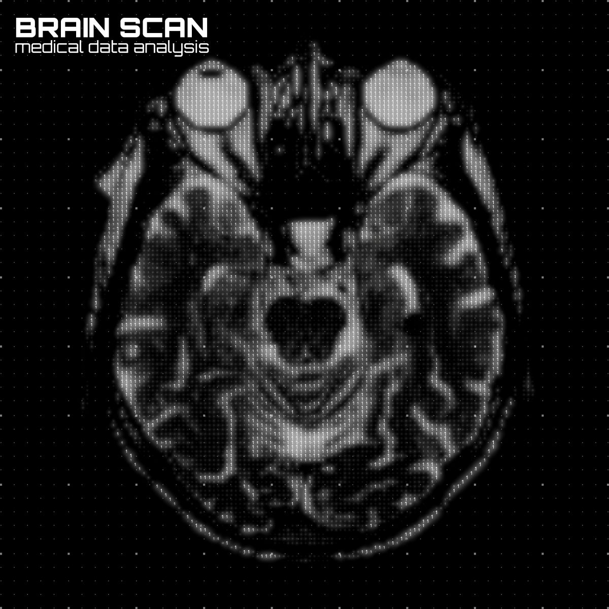
The study enrolled 20 patients who had been newly diagnosed with Parkinson’s disease, and 20 age-matched healthy subjects, to assess changes in the visual system associated with the disease. The participants underwent magnetic resonance imaging (MRI) scans, which researchers used to look at changes in the white and gray matters, as well as ophthalmologic examinations.
They found that patients presented significant changes in the brain structures associated with the visual system, such as changes in the optic radiations, decreased white matter volume and reduced volume of the optic chiasm (where the left and right optic nerves meet).
As a consequence, patients experienced visual alterations, such as an inability to perceive colors, decreased visual acuity, and a reduction in blinking, which often led to dry eyes.
According to Arrigo, these changes may appear more than a decade before the motor symptoms associated with Parkinson’s disease, which makes them potential biomarkers to diagnose and follow this disease.
“The study in depth of visual symptoms may provide sensitive markers of Parkinson’s disease,” Arrigo said. “Visual processing metrics may prove helpful in differentiating Parkinsonism disorders, following disease progression, and monitoring patient response to drug treatment.”


Kay Spitzner
Saw all of this in my mom...in hindsight...
Richard
Dry eye is diagnosed early when proper testing is done, which markers for dry eye are the important early sentinels; which are the critical ones that could early say this is definitely related to Parkinsons and what will be easily and accurately monitored if you have early sign posts for PD?
Reimund Kemmler
I have been diagnosed with mild PD back in 2013, I had to see a Neurologist ( Dr Wilcocks ) about pains in my right shoulder, as I walked into His office and before I sat down he asked me if I ever have been diagnosed with PD I replied no I haven't so he suggested that he would to some tests and I was diagnosed with Parkinson Decease.
I also have blocked tear ducts so my eyes are constantly running with tears during the day, more so in bright daylight but are dry at nights. I suffer from right mild right side tremors and have recently noticed that My right eye is dry at night.
Regina Korn
we live in New Jersey (Monroe Township} MY HUSBANDS EYE HAS BECOME STEADILY WORSE PERIFIAL VISION IS BAD. I NEED TO FIND A SPECIALIST IN NUERO/EYE PROB.
Robin Keabler
Has there ever been anyone that had asked you about a PD patient that cannot keep their eyes open, my husband could do a-lot more for himself if his eyes would stay open, he literally has to force them open or I have to tape his eyelids up and open so he can feed himself, walk or just watch tv.
Jerome
My right hand quivers and shakes sometimes and my right eye tears and the right corner of my mouth sometimes drools.