
Best Use of TMS Seen in Stimulating Two Brain Areas at High Frequency
Transcranial magnetic stimulation could help to ease patients' motor symptoms
Written by |

High-frequency stimulation of both the brain’s primary motor cortex (M1) and its dorsolateral prefrontal cortex (DLPFC) — two regions with altered activity in Parkinson’s disease — may be the optimal use of transcranial magnetic stimulation (TMS) in easing Parkinson’s motor symptoms, a meta-analysis study reported.
In analyzing data from several published studies in patients, the researchers found that TMS was generally linked to a lessening of motor symptoms. The most significant gains, moreover, were seen by targeting these two brain regions with high stimulation frequencies.
“With the advancement of neuromodulation techniques, the individualized and precise TMS regulation on [Parkinson’s] patients is essential to improving the therapeutic effect,” the scientists wrote.

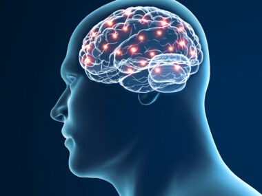
TMS uses magnetic coils placed on the skull to stimulate nerve cell activity
The study, “Comparative efficacy of transcranial magnetic stimulation on different targets in Parkinson’s disease: A Bayesian network meta-analysis,” was published in Frontiers in Aging Neuroscience.
TMS is a non-invasive neuromodulation technique aimed at stimulating and “re-setting” certain brain regions. Magnetic coils placed on the skull are used to generate a magnetic field that stimulates nerve cells’ electrical activity in the area.
Used to treat depression, TMS has gained ground as a Parkinson’s therapy, with various studies showing that the approach can ease motor symptoms like bradykinesia (slow movement) and rigidity marking the neurodegenerative disease.
Various Parkinson’s studies have evaluated low and high frequencies of stimulation and targeted several brain regions. But a protocol for TMS that produces the maximum benefits in Parkinson’s remains unclear.
To help identify such an optimal TMS approach for Parkinson’s, a team of researchers in China systematically reviewed published studies up to December 2021 reporting the results of clinical trials into TMS’ use.
A total of 36 studies — involving 1,222 Parkinson’s patients — were included in their meta-analysis.
The trials evaluated a range of different TMS protocols, varying in the number, type, and side (unilateral and bilateral) of stimulated brain regions, as well as in the frequency of stimulation. Outcomes for each study were compared against a sham procedure or conventional disease treatment.
Unilateral stimulation involves only one side of the brain, while bilateral stimulation targets both sides.
Motor function benefits were evaluated through changes in scores using the Movement Disorder Society – Unified Parkinson’s Disease Rating Scale (MDS-UPDRS) Part 3, whose higher scores indicate worse motor symptoms.
TMS was significantly superior to a sham procedure at easing motor symptoms overall, the analysis found. In particular, protocols that used a high-frequency stimulation had a stronger effect that those that used a low frequency, and bilateral stimulation was generally better than unilateral.
Analysis favors stimulating 2 specific brain areas at high frequency
Ultimately, “high-frequency TMS targeting bilateral M1, bilateral DLPFC, and both M1 and DLPFC could significantly [ease] motor symptoms of PD [Parkinson’s disease] patients,” the researchers wrote.
The primary motor cortex and dorsolateral prefrontal cortex are brain regions showing activity changes in the brains of Parkinson’s patients. M1 is the main brain region involved in coordinating voluntary movement, while the DLPFC coordinates higher level cognitive processing and communicates with M1.
Notably, both brain regions can influence the release of dopamine, a major signaling molecule, in a brain area called the striatum. A loss of dopamine in the striatum is a hallmark of Parkinson’s disease.
The most significant improvements in motor function were observed when high-frequency stimulation was administered to both M1 and DLPFC — leading to a mean 7.6-point drop in MDS-UPDRS Part 3 scores compared with a sham procedure.
Overall, this TMS approach was estimated to have a 90.7% chance of being more effective than the other approaches.
“There are several possible explanations for why M1+DLPFC showed a better performance,” the researchers wrote, noting that reasons include greater striatal dopamine release and a re-set of the communication networks known to drive Parkinson’s symptoms.
Low frequency stimulations and targeting other brain regions did not associate with significant motor improvements. The lowest performing approach involved low-frequency stimulation of both the brain’s premotor cortex and M1.
“This research proposes the optimal TMS frequency and targets for motor symptoms of PD patients,” the study noted.
Among the study’s limitations were a lack of certain clinical information, including medication use, and patients’ age or disease stage, that could influence the results. The analysis also did not assess changes in nonmotor symptoms and TMS’ long-term effects.
As such, “large-scale and multi-center clinical trials are still irreplaceable in the future to demonstrate the relationship between different parameters and clinical outcomes,” the team concluded.

Leave a comment
Fill in the required fields to post. Your email address will not be published.