
BBSome complex guides dopamine transport; may impact Parkinson’s
Researchers employed C. elegans worm to study dopamine signaling
Written by |
A complex of proteins involved in cellular trafficking, called the BBSome, plays a role in regulating the transport of dopamine, the nerve cell signaling molecule that’s deficient in Parkinson’s disease, a study reveals.
“Given the significant medical impact of altered dopamine signaling in multiple neurobehavioral disorders, further studies of how BBSome proteins regulate the dopamine transporter may lead to new strategies for treatment,” Randy D. Blakely, PhD, senior author of the study and executive director of the Florida Atlantic University (FAU) Stiles-Nicholson Brain Institute, said in a university news release.
The study, “Forward genetic screen of the C. elegans million mutation library reveals essential, cell-autonomous contributions of BBSome proteins to dopamine signaling,” was published in the Journal of Neurochemistry.
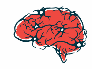
Dopamine is a neurotransmitter — a chemical that allows nerve cells to communicate with each other — that modulates various brain functions, including initiating and coordinating movement. In Parkinson’s, the nerve cells that produce dopamine, called dopaminergic neurons, are lost. This disrupts dopamine signaling and gives rise to both motor and nonmotor symptoms.
“Although intensively studied, we possess incomplete knowledge as to the networks of genes and pathways that control [dopaminergic] neuron signaling and health,” the researchers wrote.
Caenorhabditis elegans, or C. elegans, is a tiny worm used widely in research because many of its genes have the same function in humans. Both also share many common features of dopamine signaling.
“We turned to C. elegans to more efficiently elucidate the genetic, molecular, and cellular bases of neural signaling than we could with rodent models,” said Blakely, who is also the David J.S. Nicholson Distinguished Professor in Neuroscience and a professor of biomedical science in FAU’s Schmidt College of Medicine. “It turns out that the proteins involved in dopamine regulation in C. elegans are highly conserved across evolution, suggesting that lessons learned from a simpler organism with a much simpler ‘brain’ could provide clues to dopamine-linked disorders or how to better treat them.”

Dopamine signaling and swimming behavior in worms
Blakely’s team previously discovered that excessive dopamine signaling led to significant changes in worm behavior called swimming-induced paralysis, or SWIP.
“We found that an inability to constrain the actions of dopamine leads worms to freeze in a few minutes when placed in water, whereas normal worms will thrash about for up to 60 minutes or more,” Blakely said.
To identify new genes involved in dopamine signaling, the researchers used data from the Million Mutation Project (MMP), a collection of 2,007 worm strains that carry chemically induced genetic mutations. This allowed for mutant genes to be detected rapidly based on their functional impact.
A dopamine signaling blocker was given to worms in the MMP library that showed SWIP behavior. Worms that resumed swimming indicated a link between SWIP and dopamine signaling.
Researchers confirmed a connection between SWIP behavior and mutations in the gene that encodes dopamine transporter (DAT-1), a protein involved in the reuptake of dopamine into neurons after its release for signaling.
“Although, finding mutations in dat-1, a gene we already knew about didn’t accomplish our goal, this finding gave us confidence that our screen worked as intended, and that discoveries might lie ahead of us in the mutated genome of our other SWIP lines,” Blakely said.
Studying the BBSome complex
Further SWIP screening detected a mutation in the bbs-1 gene, which carries instructions for a protein that forms a complex of other proteins termed the BBSome. Overproduction of lab-made BBSsome in worms significantly rescued the SWIP defect. Moreover, SWIP in BBSome mutants was driven by excessive dopamine signaling once the animals entered the water.
In worms, the BBSome supports the formation of primary cilia, antenna-like sensory structures that enable the worms to sense their surrounding environment. In humans, the complex plays a role in transporting proteins and fat-like lipids within the cell. In fact, the rare genetic disorder known as Bardet-Biedl Syndrome (BBS) is caused by mutations that affect the BBSome.
“Our results indicate that loss of BBS-1 in worm dopamine neurons results in excess signaling by the neurotransmitter, known to inhibit movement-controlling motor neurons,” Blakely said.
Additional experiments demonstrated that bbs-1 regulated DAT-1 transport to control dopamine signaling.
“One mechanism we are considering involves a role of BBS-1 and other BBSome proteins in escorting DAT-1 encoded protein to the cell surface to keep extracellular dopamine levels low and thereby not allow a completely shutting down of movement,” Blakely added.
“We provide evidence that not only does the proper function of cilia in C. elegans [dopaminergic] neurons support normal swimming behavior, but also that bbs-1 maintains normal levels of DAT-1 trafficking,” the researchers wrote.
Leave a comment
Fill in the required fields to post. Your email address will not be published.