
Alpha-synuclein protein may travel in brain, then form toxic clumps
Florescent markers help shed light on mechanisms of Parkinson's
Written by |

The mutated form of alpha-synuclein protein — a key player in the development of Parkinson’s disease — can travel in the brain to various regions using the brain’s immune system before it builds up into toxic clumps, or aggregates, a new study has found.
The scientists suggest that these pathways contribute to the faster spread of alpha-synuclein monomers, or single alpha-synuclein protein molecules. These monomers, in turn, may function in parallel with the known propagation mechanisms of protein clumps between brain nerve cells.
Such a model varies significantly from ones that show the spread of alpha-synuclein aggregates as dependent on the clumps themselves.
“These findings suggest a mode of propagation different from that of aggregate-dependent propagation,” the researchers wrote, noting that their work suggests that “the mutant/misfolded monomer might form different aggregation species after propagation.”
The study, “Mutant α-synuclein propagates via the lymphatic system of the brain in the monomeric state,” was published in the journal Cell Reports.

Does clumping or spreading of alpha-synuclein protein happen first?
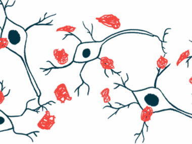
Parkinson’s disease is characterized by the dysfunction and death of nerve cells (neurons) in the brain that produce a chemical messenger called dopamine. This neurodegeneration is thought to be caused by the formation of toxic aggregates, or clusters, of mutated alpha-synuclein inside brain nerve cells.
These proteins can be transmitted to other cells in a prion-like manner. That means that protein clumps in one part of the brain can lead to the formation of clumps in connected brain areas, leading to the spread of these toxic aggregates.
However, whether aggregation or propagation — clumping or spreading —occurs first is currently unknown.
“Most experiments conducted so far only used fibrils, which are the clumps formed when monomeric [alpha]-synuclein aggregates,” Kyota Fujita, PhD, the study’s first author and an assistant professor at the Tokyo Medical and Dental University (TMDU), said in a university press release.
“The fibrils are transmitted from neurons to neurons, but it remains unclear whether monomers act in the same way,” Fujita said.
To further investigate how monomers and fibrils (protein clumps) move around the brain, Fujita and colleagues from TMDU injected viral particles into a region of the brain in mice to produce fluorescent mutated alpha-synuclein — whose movement was traceable in the brain.
The team used a mutated form of human alpha-synuclein called A53T, which has a high aggregation potential and has been linked with forms of familial Parkinson’s disease.
As early as two weeks after the injection, protein monomers were already present in regions distant from the site of injection, indicating a fast spreading of mutant alpha-synuclein.
Twelve months after the injection, alpha-synuclein was detected in protein fibrils in these remote brain regions, indicating the protein formed clusters after it traveled in the brain and was incorporated into neurons.

Study finds propagation of alpha-synuclein protein occurs before aggregates.
Wanting to understand how this propagation occurred, the researchers analyzed the three-dimensional distribution of alpha-synuclein in the brain. The team found that the protein was located in the lymphatic system of the brain, called the glymphatic system.
This system is involved in draining and renewing the fluid from the brain. While it is responsible for the elimination of toxins, it also can also transport toxins into the brain.
According to the researchers, moreover, alpha-synuclein also was present in the extracellular matrix — the structure responsible for the maintenance of cell structure — surrounding neurons. That finding suggested that the protein is taken by the matrix before entering into neurons.
When the team investigated the aggregation state of alpha-synuclein in remote brain regions, they found that fibrils were formed after the propagation of monomers occurred.
“The propagation of [alpha]-synuclein-A53T protein in a mutant/misfolded monomeric state leads to the formation of the fibrils that characterize human [Parkinson’s disease] pathology [disease manifestations],” the researchers wrote.
The amount of alpha-synuclein aggregates and the time at which they were formed varied between brain regions, independently of the distance from the injected brain region. This follows the known susceptibility of certain brain regions, such as the substantia nigra and the brainsteam, to the accumulation of toxic alpha-synuclein clumps.
“Further analyses will be necessary to confirm the fast-track brain lymphatic propagation of mutant/misfolded monomeric forms of these proteins that could occur in parallel with the neuron-to-neuron propagation of aggregated forms,” the researchers wrote.
According to the scientists, the brain lymphatic system-mediated protein propagation could be generalized to other proteins causing neurodegenerative diseases.
One key limitation of the study, per researchers, was that it was only conducted in mice. Further studies are needed to demonstrate that the spread of protein monomers via the brain lymphatic system also occurs in humans, the team said.


Leave a comment
Fill in the required fields to post. Your email address will not be published.