Alpha-synuclein blood test may aid in Parkinson’s diagnosis: Study
Test detects protein clumps in tiny, molecule-carrying sacs
Written by |

Researchers have developed a method to detect clumps of alpha-synuclein protein, a hallmark of Parkinson’s disease, within extracellular vesicles (EVs) purified from blood, providing a possible aid to Parkinson’s diagnosis before symptoms appear.
EVs are tiny membrane-bound sacs released from cells that can carry a variety of molecules, including alpha-synuclein, from one cell to another. Because EVs are scarce in biofluids and are difficult to purify, accurately measuring proteins inside EVs has been a significant challenge.
“Our recent work is providing a solution to help fill this technological gap, and gets us closer to being able [use EVs] as rich sources for clinical biomarkers,” David Walt, PhD, study lead at the Wyss Institute at Harvard University and Brigham and Women’s Hospital in Boston, said in a university news story.
The advance was detailed in the study, “Measurement of α-synuclein as protein cargo in plasma extracellular vesicles,” published in PNAS.
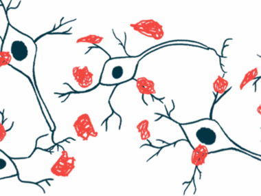
In Parkinson’s, the progressive loss of nerve cells that make dopamine, a signaling molecule the cells use to communicate, leads to various motor and nonmotor symptoms. While the cause of nerve cell death is unclear, the formation of alpha-synuclein protein clumps, known as Lewy bodies, is thought to play a role.
Lack of diagnostic tests hampers early treatment
Because brain disorders like Parkinson’s start to develop long before patients experience their first symptoms, starting treatment as early as possible, even before signs emerge, may help slow or halt disease progression. Since there are no specific tests to detect Parkinson’s disease before symptom onset, diagnosis largely relies on the manifestation of disease symptoms.
EVs are tiny sacs that can carry various biologically active molecules, including proteins, lipids (fats), and RNA, from one cell to another to facilitate cell-to-cell communication. In Parkinson’s, EVs are suspected of spreading alpha-synuclein clumps among brain cells, and therefore may carry biomarkers that predict the onset of Parkinson’s and other brain disorders.
Measuring EV cargo proteins has been challenging because EVs are present at low levels. And current methods to purify EVs by separating them from free proteins are imperfect, making it difficult to know whether a measured protein is truly inside EVs.
“The isolation of pure tissue-specific EVs from body fluids like blood or the cerebrospinal fluid surrounding the central nervous system, including the brain, and validating and quantifying their true contents with precise measurements still present formidable technical challenges,” Walt said.
Walt and colleagues developed methods to measure proteins inside EVs relative to proteins outside in blood.
Inside EVs
The team recovered EVs from biofluids using a separation technique known as size exclusion chromatography. EVs were then subjected to enzyme treatment to remove all surface-bound proteins, ensuring a purified EV population without contamination.
“We use an enzyme to gently but efficiently chew all proteins off the surface of isolated EVs, while leaving the membrane-enclosed EV interior intact,” said study co-first author Dima Ter-Ovanesyan, PhD, a senior scientist at the Wyss Institute.
To measure proteins inside EVs, researchers used an ultra-sensitive single-molecule array (Simoa) test, which allowed them to count single-protein molecules associated with EVs with specific antibodies.
When applied, most of the alpha-synuclein protein in EVs was protected and represented less than 5% of total blood alpha-synuclein. Simoa could not only measure normal, unmodified alpha-synuclein, but also detect phosphorylated alpha-synuclein, a version of the protein with a tendency to form clumps.
The team applied the methods to measure total and phosphorylated alpha-synuclein inside EVs from samples collected from people with Parkinson’s, Lewy body dementia, and healthy controls. Data showed that the ratio of phosphorylated protein relative to total protein was two to three times higher inside EVs than outside in patient samples.
“This was extremely exciting because it suggests that EVs may protect the phosphorylation state of proteins from circulating [enzymes] that would otherwise erase this highly informative mark,” said co-first author Tal Gilboa, PhD, a postdoctoral fellow at Wyss.
Walt’s team will now explore whether these tests could distinguish between people with and without Parkinson’s.
“The work by David Walt’s team presents a technological tour-de-force that brings us closer and closer to a next-generation diagnostic platform with extraordinary potential,” said Donald Ingber, MD, PhD, Wyss founding director and professor of vascular biology at Harvard Medical School and Boston Children’s Hospital. “At this point, we are not far from using these extremely rich and telling cell-derived vesicles as a window to peek into the brains of patients without requiring surgery.”
The research was supported by the Michael J. Fox Foundation, Chan Zuckerberg Initiative NeuroDegeneration Challenge Network, and Good Ventures.


