
Study links Parkinson’s hallucinations to low brain chemical activity
Patients have reduced activity in acetylcholine nerve cell network
Written by |

- Parkinson’s patients experiencing visual hallucinations show reduced activity in the brain’s acetylcholine-using nerve cell network.
- Impaired early cholinergic signaling in specific brain regions can predict which patients will later develop visual hallucinations.
- Measuring cholinergic activity may serve as a biomarker for risk and a target for future therapeutic interventions.
People with Parkinson’s disease who experience visual hallucinations have reduced activity in the brain’s cholinergic system, a network of nerve cells that use the chemical messenger acetylcholine, a brain imaging study found.
Impaired cholinergic signaling in certain brain regions early on predicted who would later develop visual hallucinations.
The researchers said measuring cholinergic activity in certain brain regions could serve as a biomarker to identify patients at a higher risk of developing hallucinations, or as a target for future treatments aimed at reducing or preventing them.
The study, “Cholinergic deficits and hallucinations in Parkinson disease,” was published in npj Parkinson’s Disease.

Measuring activity
Visual hallucinations, a non-motor symptom of Parkinson’s disease, can range in severity from minor illusions or a sense of a presence to fully formed images that persist and feel real. They are frequently linked to disease progression and/or medication side effects.
Scientists have long suspected that such hallucinations are related to problems with the brain’s cholinergic system. That’s because medications that block acetylcholine increase the frequency of hallucinations, while those that boost acetylcholine activity possibly reduce them.
However, few studies have directly measured cholinergic activity in the living brain and how it relates to the development and severity of hallucinations in Parkinson’s patients.
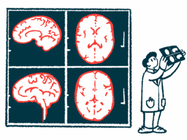
To address this gap, scientists at Washington University in St. Louis measured cholinergic activity in specific brain regions of 95 people with Parkinson’s. The team used positron emission tomography (PET) scans with a radioactive tracer called [18F]VAT, which binds to a protein involved in acetylcholine release, to detect activity.
During interviews at the study’s start (baseline), 22 participants had fully formed hallucinations, and 14 had minor or non-visual hallucinations (fleeting images or a sense of someone nearby). Among the 40 participants who didn’t have baseline hallucinations, 17 went on to develop fully formed hallucinations by a follow-up visit two to six years later.
At baseline, PET scans revealed that those with visual hallucinations had lower [18F]VAT binding, meaning less cholinergic activity, than those without hallucinations in several brain regions. These differences remained significant even after accounting for age, sex, and the duration of participants’ motor symptoms.
Affected brain regions included the thalamus, the brain’s primary relay station, which is smaller in Parkinson’s patients with more severe non-motor symptoms. Other impaired regions included the lateral occipital cortex and the middle temporal cortex, both of which are involved in complex visual processing.
Worse hallucination severity, as assessed by a self-reported questionnaire, also correlated with lower cholinergic activity in the same brain regions, as well as the cuneus, lingual gyrus, and inferior parietal cortex. The brain uses these additional interconnected brain regions for processing visual information, visuospatial attention, and spatial awareness.
“Cholinergic binding in these regions may serve as a prognostic biomarker for [visual hallucinations] in [Parkinson’s] or be promising targets for localized treatment interventions,” the team wrote.
The researchers also found that lower cholinergic activity could be detected before hallucinations began. That is, those who had no baseline hallucinations but later developed fully formed visual hallucinations already had reduced cholinergic activity at the study’s start in certain brain regions compared with patients who remained hallucination-free.
The results didn’t change when patients with dementia were excluded from the analysis. And nearly all regions remained significant after controlling for scores on the Montreal Cognitive Assessment (MoCA), which assesses early memory and thinking problems.
“[Parkinson’s disease] participants with full-formed [visual hallucinations] showed less cholinergic innervation of widespread [brain] regions,” the researchers wrote. “The majority of these deficits correlated with increased hallucination severity and predicted the future development of [visual hallucinations].”

Jose Garcia
Can you reverse this deficit?