
3-D Images of Mutated Protein Variations May Increase Understanding of Parkinson’s
Written by |
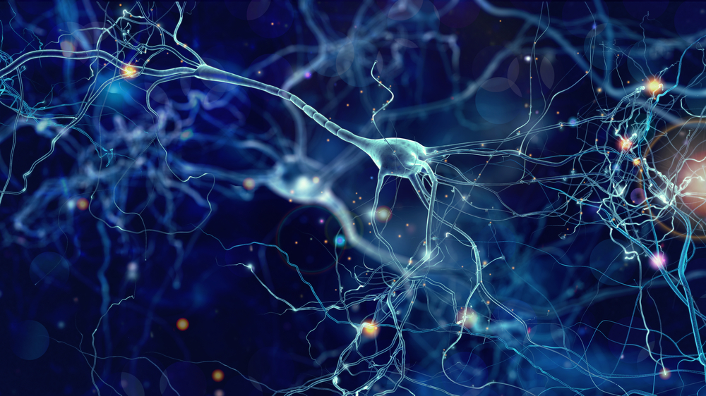
3-D images of 11 mutant versions of a protein connected with Parkinson’s disease (PD) and amyotrophic lateral sclerosis (ALS) are helping scientists understand how the mutations changed the structure of the protein and damaged its functioning.
The study, “Structural insights into human angiogenin (ANG) variants implicated in Parkinson’s disease and Amyotrophic Lateral Sclerosis,” was published in Scientific Reports.
University of Bath researchers studied a protein in the central nervous system known as angiogenin, or ANG. It is believed to have both neuroprotective and regenerative functions, protecting cells in the brain and spinal cord. ANG mutations have been implicated in the development of Parkinson’s and ALS.
The research team, which included cell and structural biologists, used software designed to produce 3-D molecular models to create ANG mutation models.
The images give them another tool for studying how changes in molecular structure impair ANG’s functioning. The mutations are believed to contribute to disease progression in both Parkinson’s and ALS.
In previous studies, the team documented the effects that malfunctioning ANG variations had on embryonic stem cells. The mutations prevented ANG from reaching the cells’ nuclei, impairing the protein’s ability to function, they discovered. In addition, they found less ANG in neurodegenerative diseases such as Parkinson’s and ALS.
“We have extended our structural study to eleven new human ANG PD/ALS variants,” the team wrote. They added that they had also created “detailed molecular features of these variants in comparison with the normal ANG protein, thus progressing from understanding structure to using structure to understanding disease.”
Refining the molecular-mutation structures they created by adding more detail could lead to an even better understanding of the way Parkinson’s and ALS development, the team said.
“We hope that the scientific community can use this new knowledge to help design new drugs that will bind selectively to the defective protein to protect the body from its damaging effects,” Professor K. Ravi Acharya, the study’s senior author, said in a news release. He is a structural biologist in the University of Bath’s Department of Biology & Biochemistry.
The team said it hopes the structures it created will lead to an understanding of “the biological roles for these variants which have been implicated in PD and ALS.”


Leave a comment
Fill in the required fields to post. Your email address will not be published.