
Researchers discover new cellular mechanisms in Parkinson’s disease
Mazzulli: 'Other proteins and RNA inclusions' besides alpha-synuclein
Written by |

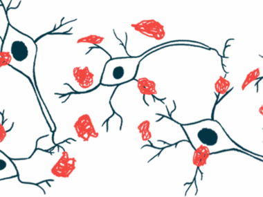
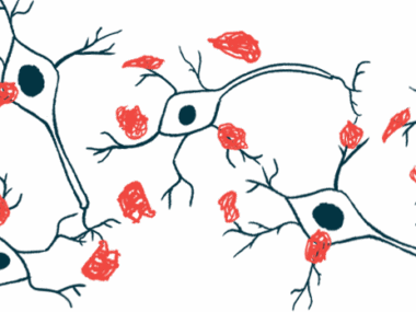
Clumps of RNA-binding proteins have been discovered in the nucleus of nerve cells derived from people with Parkinson’s disease, according to a new study.
Generated by a “self-propagating” cycle, these clumps were different and separate from those made from the protein alpha-synuclein, a well-known feature of Parkinson’s.
A second study by the same research team showed that a metabolic pathway that plays an essential role in protein folding was disrupted in cells derived from Parkinson’s patients. These defects also resulted in protein clumps forming in nerve cells.
“When you think about Parkinson’s treatments, anything that’s in the preclinical stage now or in clinical trials is targeting one pathway,” Joseph Mazzulli, PhD, the lead author of both studies and associate professor at the Northwestern University Feinberg School of Medicine in Illinois, said in a university news release. “Our studies show that multiple pathways are perturbed in the disease and so we think that rescuing multiple pathways at once is essential for disease modification.”

Two more aggregating proteins
The first study, “Nuclear aggregates of NONO/SFPQ and A-to-I-edited RNA in Parkinson’s disease and dementia with Lewy bodies,” was published in Neuron.
In neurodegenerative diseases such as Parkinson’s and Lewy body dementia (LBD), alpha-synuclein becomes misfolded and aggregates into large clumps, called Lewy bodies, which is thought to disrupt the function of neurons.
In previous work, Mazzulli and his colleagues showed that alpha-synuclein-induced deficits led to proteins building up in the endoplasmic reticulum (ER), the cellular structure involved in protein production, folding, and transport. As a result, the function of lysosomes, the cell’s recycling centers, was obstructed, causing protein aggregation.
Here, Mazzulli’s team used neurons derived from people with familial Parkinson’s and LBD to identify the proteins susceptible to aggregation in neurons affected by Parkinson’s.
Unexpectedly, the team found aggregates inside the cell nucleus composed of two proteins known to bind RNA: NONO and SFPQ. Alpha-synuclein aggregates are typically found outside the nucleus, but these new aggregates were separate from Lewy bodies and accumulated at levels comparable to alpha-synuclein. RNA is a molecule that helps carry genetic information from DNA to create proteins and is involved in various cellular processes, acting as a messenger between DNA and proteins.
Aggregated NONO and SFPQ were found to reduce the activity of a gene that encodes for the protein ADAR3. Known as an RNA editing inhibitor, ADAR3 blocks an enzyme that edits RNA by converting adenosine (A), an RNA building block, into inosine (I), a molecule not typically found in RNA.
Without ADAR3, so-called A-to-I RNA editing increased, destabilizing RNA and causing it to accumulate abnormally in the nucleus. In turn, inosine-containing RNAs tightly bound to SFPQ and triggered SFPQ aggregation in dopamine-producing neurons, the nerve cells lost in Parkinson’s. This process resulted in a “self-propagating” disease-driving cycle.
RNAs affected included those from axons (nerve fibers), synapses, the gaps where communication between neurons occurs, and mitochondria, the cell components that generate most of its energy.
“The field has been really focused on alpha-synuclein aggregation,” Mazzulli said. “ We’re not saying that’s not important, but we’re showing that there are other proteins and RNA inclusions involved in Parkinson’s pathogenesis that we haven’t seen before.”
Disrupted metabolic pathway
Mazzulli’s second study, “The hexosamine biosynthetic pathway rescues lysosomal dysfunction in Parkinson’s disease patient iPSC derived midbrain neurons,” published in Nature Communications, investigated the connection between glucose metabolism and protein misfolding in neurons affected by Parkinson’s.
Altered glucose metabolism and protein misfolding are key characteristics of age-related neurodegenerative diseases, such as Parkinson’s. Little is known about the mechanism that links these two disease-related features, however.
Again using neurons derived from Parkinson’s patients, the researchers discovered a disruption in the hexosamine metabolic pathway, which is important for protein production, folding, and transport in the cell’s ER.
The disruption of this pathway reduced the production of N-linked glycans, modified carbohydrates that support protein folding in the ER. At the same time, levels of glucose and other precursor molecules increased. Without proper N-linked glycan production, proteins within lysosomes misfolded and accumulated.
The source of the metabolic disruption was the depletion of GFPT2, an enzyme that provides the precursors for producing N-linked glycans.
“We found a metabolic enzyme in the hexosamine pathway that was depleted, called GFPT2. It’s an important enzyme and provides the precursors for N-glycosylation,” Mazzulli said. ”If you wipe that out, protein folding in the [ER] is severely compromised, leading to proteome stress.”
Increasing the production of GFPT2, or treating cells with N-acetylglucosamine, a molecule in the hexosamine pathway, improved protein folding in the ER, rescued lysosomal function, and reduced alpha-synuclein aggregation.
“Our data indicate that the hexosamine pathway integrates glucose metabolism with lysosomal activity,” the researchers wrote. “These findings offer new methods to restore [protein balance] by hexosamine pathway enhancement.”

Leave a comment
Fill in the required fields to post. Your email address will not be published.