
New Brain Pathway Key to Movement Found in PD Patients, Study Reports
Written by |
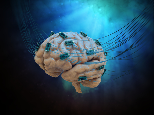
Placing electrodes directly in the brains of people with Parkinson’s disease revealed a “hyperdirect” pathway between two regions of the brain responsible for movement and cognition, a study reported.
This pathway was shown to be important in being able to stop body movements once initiated. Modulating or controlling this pathway may be a therapeutic strategy for treating movement disorders such as Parkinson’s.
The study, “Prefrontal-Subthalamic Hyperdirect Pathway Modulates Movement Inhibition in Humans,” was published in the journal Neuron.
Parkinson’s disease is characterized by a progressive loss of coordination and movement leading to involuntary tremors and other motor symptoms.
Stopping a body movement that has already been initiated is important for motor control, which is thought to be mediated by a pathway between two regions in the brain. The pathway connects the subthalamic nucleus (STN), which is involved in many complex motor and non-motor functions, and the inferior frontal gyrus (IFG), associated with cognition.
“This pathway is critical to controlling movement overall,” Witney Chen, a graduate student at the University of California, San Francisco (UCSF) and the study’s first author, said in a news story.
“We’re interested in understanding how the brain controls the ways we can stop movement because when this control isn’t functioning properly, it can result in movement disorders such as Parkinson’s,” Chen said.
“This is more than just being able to quickly stop your step into the street if you see oncoming traffic,” she added.
Evidence suggested that the STN-IFG pathway exists in animals. However, only indirect imaging studies have supported its importance in humans, and in the workings of Parkinson’s disease.
To explore this pathway, Chen and colleagues based at UCSF designed a study involving 21 Parkinson’s patients in which electrodes were placed directly in the brain in both the IFG and STN regions. The goal was to gather as much information as possible about this pathway in humans.
“It’s a wonderful opportunity to study the human brain as an intact system,” said Philip Starr, MD, PhD, co-director of the UCSF Surgical Movement Disorders Center and the study’s senior author. “And fortunately, Parkinson’s patients are especially eager to volunteer. They’re often people who had normal lives for a long time, and now they have this disorder and they really want to contribute to understanding and treating it.”
“These experiments can really only be done well with invasive electrodes at both ends of the pathway,” Starr added.
The Parkinson’s patients enrolled in the study were already scheduled to have electrodes implanted in the STN region of the brain, a standard procedure for deep brain stimulation (DBS), often used on those with mid-stage disease. As there are no pain receptors in the brain, the participants are awake during surgery and can confirm the placement and function of the DBS implants with physicians.
During the procedure, electrodes also were placed on the surface of the brain, about five centimeters (2 inches) from the DBS implants. Chen noted that these electrode could easily be removed after the experiments.
The team then recorded high-resolution electrical impulses focusing on location and time. They found the response to STN stimulation was detected very quickly (low latency) in the IFG region, which demonstrated a “hyperdirect” connection between these two parts of the brain.
A second experiment was conducted to measure the ability to stop a body movement. Here, patients were shown either a right or left arrow on a screen as a “go” signal, to which they responded by pushing a respective right or left button in response. Randomly, they received a “stop” signal after the “go” signal and the time taken to stop movement was measured.
The results showed that the longer the IFG and STN signals were simultaneously activated — representing a higher synchronization between both regions — the faster the participants stopped their action, and that faster initiation of activity was found to be important for successful stopping. These findings, shown across all participants tested, demonstrated a direct synchronization between these two brain regions in movement control.
Although movement inhibition has been found to be impaired in people with Parkinson’s, the team did not find physiological factors or stopping behaviors to be associated with parkinsonian severity, as assessed by the Unified Parkinson’s Disease Rating Scale.
“Our study is the first to show that the hyperdirect circuit co-modulation is linked to stopping behaviors, which has broad implications for stimulation-based therapies to treat maladaptive movement inhibition,” the scientists concluded.
“These findings may inform therapies to treat disorders featuring perturbed movement inhibition,” they added.
The next step is to study the role of the IFG and STN pathway in more real-life settings using electrodes that can record brain activity over longer periods of time, the researchers said.
“Using this technology, we can start to tease apart what this circuit is doing in real life when people are moving, talking, walking, playing music or sports or whatever they want to do,” Chen said.
“We’re really pursuing these therapeutic aspects, because we think modulation in this circuit can translate to better clinical outcomes,” she concluded.


Leave a comment
Fill in the required fields to post. Your email address will not be published.