
MRI Technique Helps Distinguish Between Parkinson’s and Progressive Supranuclear Palsy, Study Reports
Written by |
A specific magnetic resonance imaging (MRI) measure can accurately predict the development of eye movement abnormalities, helping to identify patients who develop progressive supranuclear palsy with parkinsonism (PSP-P) after an initial diagnosis of Parkinson’s, a study shows.
The study, “Refining initial diagnosis of Parkinson’s disease after follow‐up: A 4‐year prospective clinical and magnetic resonance imaging study,” appeared in the journal Movement Disorders.
A clinical diagnosis of Parkinson’s may be inaccurate in the early stages of the disease, when its characteristic motor symptoms are not fully manifested. PSP-P has a similar clinical presentation to idiopathic Parkinson’s (of unknown cause), including bradykinesia (slowness of movement), limb rigidity, and tremor, leading to difficulty in differentiating between the two.
Recent diagnostic criteria defined probable PSP-P as vertical gaze abnormalities (VGA) — difficulty in moving the eyes up and/or down — associated with levodopa‐resistant parkinsonism, a term for neurological disorders that cause movement problems similar to those of Parkinson’s. However, patients with PSP-P may never develop VGA, meaning the prevalence of this disorder could be underestimated.
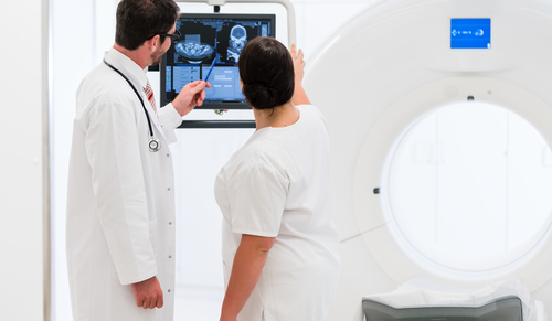
MRI has been a helpful tool to diagnose PSP and to predict the appearance of gaze abnormalities in people with PSP-P. A new version of the Magnetic Resonance Parkinsonism Index (MRPI), named MRPI 2.0, has shown high accuracy in distinguishing cases of Parkinson’s from those of PSP-P. The MRPI can be used in MRI studies to predict the presence of PSP in patients with clinically unclassifiable parkinsonism.
The Parkinson’s Disease News Today forums are a place to connect with other patients, share tips and talk about the latest research. Check them out today!
However, studies on the clinical features of PSP-P in patients initially diagnosed with Parkinson’s are still lacking.
To address this, researchers in Italy followed a group of 110 individuals — 73 of whom were men, with a mean age at examination of 62.9 years, and a mean disease duration of 4.4 years — with probable or possible Parkinson’s and 74 healthy individuals used as controls over four years.
The investigators conducted annual clinical evaluations to assess the appearance of VGA without early postural instability, which strongly suggests PSP‐P. MRI scans were performed at the beginning of the study and at the end of the follow-up period.
They also evaluated whether MRPI 2.0 helped predict the development of PSP-P in patients initially diagnosed with Parkinson’s.
At the start, 21 of 40 individuals with possible Parkinson’s and all 70 individuals with probable Parkinson’s were responsive to levodopa. Of all the patients, 100 retained their initial diagnosis, and 10 (9.1%) developed VGA and had their diagnosis changed to PSP-P, nine of whom only showed a moderate response to levodopa.
Specifically, all 10 patients whose diagnosis changed showed slowness of vertical saccades during follow-up, which refers to quick, simultaneous movements of both eyes that abruptly change the point of fixation. Vertical supranuclear gaze palsy (resulting from a cerebral impairment) in five patients was associated with higher imaging biomarkers values than slowness in vertical saccades. All 10 patients had at least three years of parkinsonism without postural instability.
All MRI measures — including MRPI and MRPI 2.0 — were significantly different between patients with Parkinson’s and those at PSP-P both at the start of the study and at the end of follow-up. At the beginning, MRPI 2.0 was the most accurate (100%) biomarker in predicting the appearance of VGA, “enabling [PSP-P] patients to be identified at the earliest stage of the disease,” according to the researchers.
Although the number of patients whose diagnosis was changed is small, the researchers said that their findings “demonstrate the usefulness of these new imaging biomarkers, and specifically of the MRPI 2.0, in predicting the development of VGA and the clinical evolution towards PSP phenotypes in patients with the initial diagnosis of [Parkinson’s].”
Most clinical variables — including motor function, assessed with the Unified Parkinson’s Disease Rating Scale–Motor Examination (UPDRS‐ME), and cognitive function, measured with the Mini-Mental State Exam — also showed a marked difference between the two groups at follow-up. However, clinical variables were less accurate than imaging biomarkers in predicting VGA.
Data further showed that disease progression was more signficant in patients with PSP‐P, as assessed with MRI and UPDRS‐ME.


Nancy Turley
Could these articles be more user friendly for printing. I don't need 10 pages of only the left column printed and half of that pictures!
Would appreciate a PRINT icon at the beginning of an article that would print something that is concise.