
Parkinson’s-linked gene mutations may drive iron buildup in brain cells
Cell study links abnormal LRRK2 activity to oxidative stress and ferroptosis
Written by |
- Parkinson’s-linked LRRK2 mutations were associated with abnormal iron buildup in neurons and astrocytes in laboratory cell models.
- The mutations were also linked to oxidative stress and lipid damage associated with ferroptosis, an iron-dependent form of regulated cell death.
- Experimental LRRK2 inhibition reduced excess iron and lipid peroxidation in the cell models, pointing to iron regulation as a potential therapeutic target.
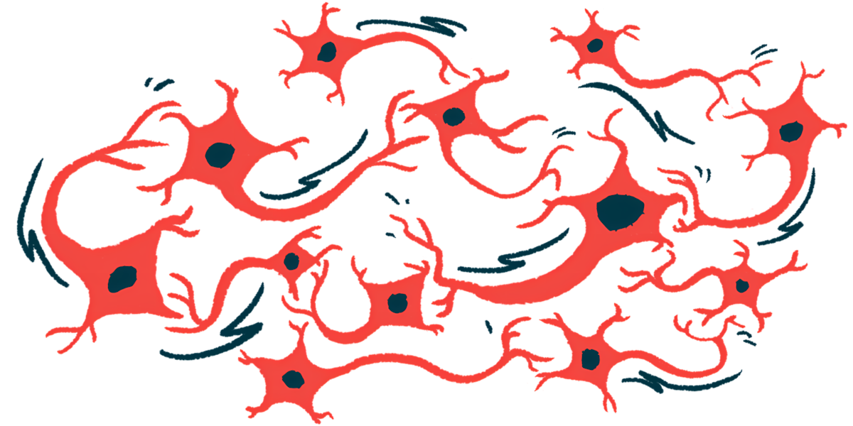
Disease-causing mutations in the LRRK2 gene may disrupt iron regulation and promote cell damage, potentially contributing to the loss of dopamine-producing nerve cells in Parkinson’s disease, a study found.
The findings suggest that abnormal LRRK2 protein activity may contribute to iron accumulation inside cells and promote oxidative stress and processes linked to ferroptosis, an iron-dependent form of regulated cell death.
“What we discovered is that mutations in the LRRK2 gene directly change the way cells handle iron,” Matthew J. LaVoie, PhD, director of the University of Florida’s Center for Translational Research in Neurodegenerative Disease and one of the researchers who led the study, said in a press release.

LRRK2 mutations may link Parkinson’s to abnormal iron buildup
The study, “Parkinson’s disease LRRK2 mutations dysregulate iron homeostasis and promote oxidative stress and ferroptosis in human neurons and astrocytes,” was published in Molecular Neurodegeneration.
Parkinson’s disease is caused by the progressive loss of dopaminergic neurons, nerve cells that produce dopamine, a signaling molecule involved in motor control. These neurons are primarily found in a brain region called the substantia nigra.
Iron accumulation is thought to contribute to neuronal damage by promoting oxidative stress, an imbalance between harmful free radicals and the body’s antioxidant defenses, and ferroptosis. Mutations in LRRK2, among the most common genetic causes of Parkinson’s, have previously been linked to higher levels of iron in the substantia nigra.
However, how LRRK2 mutations affect iron regulation in human neurons and astrocytes, nerve-supporting cells that help regulate iron availability, and whether this contributes to neuronal damage, remain poorly understood.
“For the longest time, we’ve seen a correlation in living patients between iron deposition in affected regions of the brain and Parkinson’s disease, but we couldn’t understand it,” said study author Adam Mamais, PhD, a research assistant professor of neurology.
To investigate these questions, researchers used both patient-derived and gene-edited human induced pluripotent stem cell (iPSC) models carrying disease-causing LRRK2 mutations, including G2019S, R1441C, R1441G, and Y1699C, to study their effects on iron regulation. iPSCs are made by reprogramming adult cells into a stem cell-like state, after which they can be guided to develop into different cell types.
Mutant cells show excess iron and altered iron storage
Compared with control iPSCs without disease-causing LRRK2 mutations, mutant iPSC lines generally had higher levels of labile iron, a form of iron that is readily available for use by cells but can become harmful when it accumulates, as well as higher levels of ferritin, a protein that stores iron. Cells carrying the R1441C mutation also had higher total iron levels.
Higher levels of labile iron were also present in neurons derived from iPSCs carrying the R1441C or G2019S mutations.
Treatment with MLi-2, an experimental LRRK2 inhibitor, reduced excess labile iron in iPSCs carrying the G2019S mutation and partially reduced it in those with the R1441C mutation, suggesting that abnormal LRRK2 activity may contribute to iron accumulation.
The researchers next looked specifically at lysosomes, cellular structures that help break down and recycle materials while also helping regulate iron inside cells. They found higher levels of lysosomal iron in iPSCs carrying LRRK2 mutations than in cells without the mutations. The same increase was seen in neurons and astrocytes derived from these cells.
MLi-2 treatment also reduced iron buildup inside lysosomes in astrocytes carrying each of the three mutations tested — R1441C, Y1699C, and G2019S. The effect was strongest in cells with the G2019S mutation, where iron levels returned to those seen in cells without the mutation.
Cells with the G2019S mutation also had higher levels of the two ferritin components measured, while R1441C cells showed a trend toward lower levels of one of them. Changes were also seen in proteins involved in sensing and controlling iron levels, particularly in G2019S cells.
“What we’ve discovered is a mechanism through which iron is dysregulated, and it is corrected by inhibitors of LRRK2, which are under clinical development for treatment of Parkinson’s disease,” LaVoie said.
Rab8a signaling may help explain iron buildup
Experiments in iPSCs and iPSC-derived neurons showed that knocking out Rab8a, a protein regulated by LRRK2, increased labile iron, a readily available form of iron inside cells. Restoring Rab8a in Rab8a-deficient iPSCs brought iron levels closer to normal, suggesting that disrupted LRRK2-Rab8a signaling may contribute to iron buildup.
Neurons carrying LRRK2 mutations had increased oxidative stress and lipid peroxidation, a type of damage to fatty molecules that are important components of cell membranes. Both effects were reduced when the cells were treated with deferoxamine, a compound that binds excess iron.
MLi-2 reduced lipid peroxidation, bringing levels closer to those seen in neurons without the mutations.
“What our paper shows is that Parkinson’s disease is beyond aggregated protein,” LaVoie said. “We see that mutations in the most common genetic type of late-onset Parkinson’s cause mismanagement of iron in different brain cells.”
According to the researchers, these findings point to iron dysregulation as a potential therapeutic target in LRRK2-associated Parkinson’s disease. Because the study was conducted in lab-grown cell models, more research is needed to determine whether targeting specific iron-handling pathways could help alter disease progression.



gary canner
What does this portend for the PWP who has LRRk2 mutation AND TAKES IRON DAILY TO BOOST HIS FERRITIN/IRON COUNT TO COUNTER EXCESSIVE EXHAUSTON?