Levels of fat molecules altered in brains of Parkinson’s patients: Study
Written by |
People with Parkinson’s disease have altered levels of certain fat molecules, known as lipids, in their brains, and this may play a role in disease development and progression, a new study from the U.S. reports.
The researchers used postmortem brain tissue samples to analyze changes in lipids in 40 Parkinson’s patients and 43 age- and sex-match individuals without the neurodegenerative disease.
According to the team, “this is the first prospective study to employ [this type of] cutting-edge … approach to analyze a vast array of … lipid species” in the brains of people who have died from Parkinson’s.
“These findings highlight lipid dysregulation in PD [Parkinson’s disease] and point to potential biomarkers for diagnosis, warranting further validation,” the researchers wrote. The team stressed that “the observed associations may not imply causation,” and noted that more research is needed.
The study, “Lipid profiling of Parkinson’s disease brain highlights disruption in Lysophosphatidylcholines, and triacylglycerol metabolism,” was published in the journal npj Parkinson’s Disease.
Fat molecules, or lipids, play a range of vital roles in the body, from being a useful source of energy to helping form the membranes of cells. In this study, a team led by scientists at the Corewell Health Research Institute in Royal Oak, Michigan, set out to examine how different lipids are altered in the brains of people with Parkinson’s.
“Lipids are involved in many aspects of PD [disease biology] … and lipid dysregulation or disruptions in lipid [balance] could contribute to the [disease’s development],” the team wrote.
Brain tissue samples studied from people with, without Parkinson’s
The scientists collected the postmortem tissue samples, then conducted detailed assessments of lipid contents in a specific area of the brain known as the primary motor cortex. This region helps regulate motor function and cognition.
From hundreds of lipids analyzed, the researchers identified 95 that were present at significantly altered levels in the brains of people with Parkinson’s versus healthy controls. Among the altered lipids, the researchers noted that Parkinson’s patients had abnormally high levels of triacylglycerols (TAGs) but abnormally low levels of lysophosphatidylcholines (LPCs).
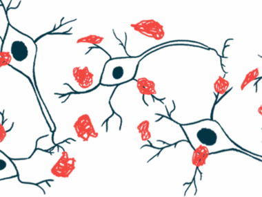
TAGs are fatty molecules used for energy storage. Importantly, they are known to interact with alpha-synuclein, a protein that characteristically forms toxic clumps in the brain cells of people with Parkinson’s.
“Elevated TAGs in PD brains could indicate an abnormal accumulation of lipids, which might be related to altered energy metabolism or impaired lipid processing in the brain,” the researchers wrote.
Overall, these [lipid] changes could contribute to the [development and progression of Parkinson’s disease], potentially affecting neuronal function, membrane integrity, and inflammatory responses.
LPCs, for their part, are involved in maintaining the structure of cell membranes. The researchers speculated that low LPC levels could lead to disruptions in the structures of nerve cell membranes, which could contribute to nerve damage in Parkinson’s.
“Overall, these [lipid] changes could contribute to the [development and progression of Parkinson’s disease], potentially affecting neuronal function, membrane integrity, and inflammatory responses,” the researchers wrote.
According to the team, these findings “could conceivably have implications for the development of biomarkers that could facilitate early detection, risk assessment, and therapeutic monitoring” in Parkinson’s.
Abnormalities in fat molecules differ between males and females
In further statistical analyses, the researchers noted that specific patterns of lipid abnormalities were significantly different between male and female Parkinson’s patients.
This is important, the scientists noted, because Parkinson’s predominantly affects males for reasons that remain incompletely understood. The team suggested that these differences in lipid profiles could play a role in sex-specific differences in Parkinson’s.
“Identifying sex-related lipid classes and species signatures that can be used as sex-specific [Parkinson’s] markers … are essential for personalized medicine strategies,” the researchers wrote.
The scientists also used machine learning models to see how well lipid profiles could distinguish between people with or without Parkinson’s. These results indicated that lipid analysis could accurately identify about more than 75% of Parkinson’s patients and more than 85% of those without Parkinson’s.
The scientists stressed that this study was limited to samples from a few dozen people, and that the analyses only used brain samples collected after death. The team emphasized a need for further work to validate these findings and investigate the clinical implications of lipid alterations in Parkinson’s disease.
“Further research would be needed to explore the exact mechanisms by which these lipid alterations contribute to disease progression and whether they might serve as potential biomarkers or therapeutic targets,” the scientists concluded.


