How Parkinson’s Disease Spreads in the Brain Shown in Mouse Study
Alpha-synuclein clumps may move from neuron to neuron in 'chain reaction' via lysosomes
Written by |

Clumps of alpha-synuclein protein, which build to toxic levels in the brain and spinal cord of Parkinson’s disease patients, spread via a cellular waste-ejection process, according to a study in mice.
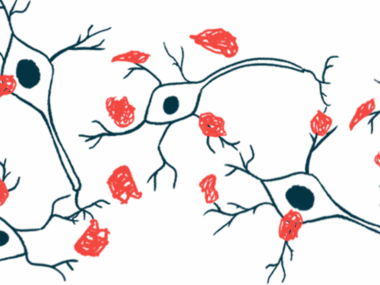
These clumps, which failed to be broken down in lysosomes, the cells’ recycling centers, are released by nerve cells in the brain through a process called lysosomal exocytosis. As a result, these alpha-synuclein aggregates spread to neighboring nerve cells, triggering further clump formation and disease progression.
These findings, which resolved one of the mysteries of Parkinson’s disease, may lead to new strategies for treating or preventing the progression of the neurological disorder, the researchers noted.
“Our results also suggest that lysosomal exocytosis could be a general mechanism for the disposal of aggregated and degradation-resistant proteins from neurons — in normal, healthy circumstances and in neurodegenerative diseases,” Manu Sharma, PhD, the study’s senior author of the Weill Cornell Medicine, said in a university press release. Sharma is an assistant professor of neuroscience at Cornell’s Feil Family Brain and Mind Research Institute.
The study, “Lysosomal exocytosis releases pathogenic α-synuclein species from neurons in synucleinopathy models,” was published in Nature Communications.
Parkinson’s disease is marked by the alpha-synuclein protein aggregating to toxic levels inside nerve cells that communicate with other neurons by secreting a signaling molecule called dopamine. Progressive loss of these cells results in abnormally low levels of dopamine, leading to motor and nonmotor symptoms.
Nerve cell loss in Parkinson’s originates in one part of the brain and then spreads to other brain areas. This observation, combined with alpha-synuclein being detected in the fluid that surrounds the brain and spinal cord, suggests these toxic aggregates may spread from affected neurons to healthy neurons in an infection-like, chain reaction process.
The presence of these toxic clumps is known to attract normal alpha-synuclein toward them and as they grow larger they break into smaller aggregates that continue to spread.
However, the molecular and cellular mechanisms underlying the spread of alpha-synuclein aggregates remain unclear, leading Sharma and his research team to evaluate whether lysosomes — sphere-shaped cellular compartments that break down unwanted or damaged molecules — were involved in this process.
Prior research has linked lysosomal abnormalities to Parkinson’s and to other neurodegenerative disorders.
The team used a mouse model that overproduces a human alpha-synuclein protein with a mutation associated with familial Parkinson’s that’s prone to form aggregates. By five months of age, these mice start to show signs of neurodegeneration.
Crossbreeding this model with mice with impaired lysosome function was found to accelerate alpha-synuclein clumps’ accumulation in brain tissue.
In 6-month-old Parkinson’s mice, disease-causing alpha-synuclein aggregates, but not single molecules, were detected inside lysosomes isolated from the animals’ brain tissue.
A sizable portion of mature aggregates, about 20%, were specifically found in nerve cells’ lysosomes, particularly within the space that contains enzymes that break down molecules. The team also confirmed that the mouse model had alpha-synuclein aggregates in the fluid surrounding the brain and spinal cord.
These clumps were not encased within membranes and further experiments revealed the release of alpha-synuclein clumps occurred by lysosomal exocytosis.
In this process, the membrane encompassing lysosomes fuses with the outer cell membrane, releasing the contents of the lysosome outside cells. Exocytosis was shown to be mediated by the SNARE protein complex, which physically joins the two membranes.
Selectively disrupting lysosomal exocytosis reduced the release of aggregates, demonstrating that “SNARE-dependent exocytosis of lysosomes is essential and rate-determining for the release of [disease-causing alpha-synuclein clumps] from neurons,” the research team wrote.
Aggregates released from the mouse model’s neurons were able to drive the formation of more clumps from single alpha-synuclein molecules, analyses showed. Again, suppressing lysosomal exocytosis significantly reduced this aggregation-promoting effect, making it a possible therapeutic approach for Parkinson’s, Sharma said.
These findings “provide evidence of the accumulation of [disease-causing alpha-synuclein clumps] in neuronal lysosomes,” and point to “lysosomal exocytosis as a pathway of release” of these aggregates outside cells, the researchers said.
“We don’t know yet, but neurons might be better off, even in the long term, if they keep these aggregates inside their lysosomes,” Sharma said. “We see a similar impairment of lysosomal function in some genetic disorders, but these don’t necessarily lead to a Parkinson’s level of disease.”
“Key gaps still remain in how the [disease-causing alpha-synuclein clumps] are targeted and trafficked to lysosomes, as well as in the fate of extracellular [alpha-synuclein] aggregates following their release from neurons,” the researchers wrote.


