Differences in Mouse, Human Astrocytes May Impact Research
Written by |

Gut inflammation model
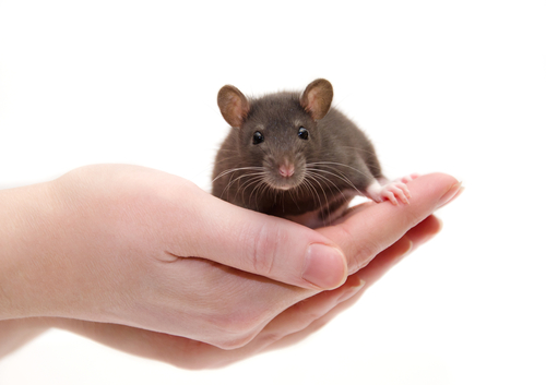
Brain cells called astrocytes behave differently under stressful conditions in humans as compared with mice, new research suggests.
Because mice are a commonly used model for research in Parkinson’s disease, these “species-specific differences” could have important implications for how such research is done, according to a team of scientists from UCLA, who led the study.
“These findings reveal important mechanistic differences between human and mouse astrocytes and provide insight into how mouse models of neurodegeneration and stroke can be improved to achieve better translation to humans,” the investigators wrote.
The study, “Conservation and divergence of vulnerability and responses to stressors between human and mouse astrocytes,” was published in Nature Communications.
The biggest problem with animal studies, according to the investigators, is that many mouse models of neurodegenerative disorders show milder nerve cell degeneration characteristics as compared with human patients. Also at issue for research is that mouse models in some cases can fully recover function, while human patients have irreversible damage.
“These limitations represent a key barrier in translational research, as over 90% of neurological drug candidates with promising animal data failing in human clinical trials,” the team wrote.
Astrocytes, which get their name from their star-like shape, are cells found in abundance in the nervous system. Unlike neurons (nerve cells), astrocytes don’t send electrical signals. But they play many important functions in supporting neuronal function and health — many of which are only beginning to be understood.
The majority of research into astrocyte biology has been done using mouse cells. While mice are a useful research organism in many regards, they also are very different from people, biologically, with differences in body size, life span, ecological niche, and behavior.
Understanding these differences is crucial for translating findings from mice to people, which has historically posed a massive hurdle in drug development. In fact, more than nine out of every 10 treatments for neurological disorders that show promise in preclinical studies don’t get approved.
Now, the team from UCLA and their colleagues sought to better characterize the differences between developing astrocytes from mice or humans. They used a specially developed technique to keep the astrocytes alive and healthy in a laboratory setting.
First, the researchers examined the gene expression profiles of astrocytes from the different species. Gene expression is essentially defined as which genes within a cell are “turned on or off, and by how much.” By looking at which genes are more or less active, researchers can look for patterns that provide clues to the overall activity of the cell.
The gene profiles of astrocytes from mice and humans were largely similar — which was expected, since they’re the same kind of cell. Nonetheless, more than 8,000 genes were expressed at significantly different levels in the two species, the investigators found. Very generally, there was greater activation of metabolism-related genes in mouse astrocytes, whereas human astrocytes had more activation of genes related to inflammation. Additional cellular experiments also suggested metabolic differences between the two species.
The researchers also evaluated the gene expression profiles of human astrocytes that were grown in the brains of living mice. These cells’ profiles were similar to those of other human cells, which demonstrates that the difference between species is intrinsic to the astrocytes themselves and not due to other differences in the species’ brains.
Next, the team examined how the astrocytes changed in response to various threats and damages, such as simulated viral infection, inflammation, low oxygen levels, and oxidative stress (highly reactive molecules that can damage cellular structures).
Compared with mouse cells, human astrocytes were much more vulnerable to damage by oxidative stress. A more detailed examination revealed that this difference likely has to do with differences in activity in the cells’ mitochondria, the cellular structures mainly responsible for producing energy.
“Mitochondria from mouse astrocytes are highly resilient to oxidative damage … In contrast, mitochondria from human astrocytes are quickly damaged and cannot keep up with the cellular energy demand when exposed to oxidative stress,” the researchers wrote.
Other differences, such as high levels of detoxifying enzymes, also were noted, and likely inform the cells’ differing responses to oxidative stress. Of note, oxidative stress is increased in Parkinson’s and other neurological diseases.
The scientists suggested that engineering mice so that their astrocytes are more susceptible to oxidative stress might make them more suitable models for research into neurological diseases.
Another noteworthy finding from the study is that mouse astrocytes — but not human cells — activate a cellular program that aims to support neuron health in response to low oxygen levels (hypoxia). The investigators suggested that this finding may be especially relevant for research in stroke. They also speculated that further research might seek to determine how a similar program could be turned on in human cells.
“Investigating how signal transduction occurs in mouse astrocytes that links hypoxia to neuronal growth genes may lead to therapies that activate the pro-neuronal growth program in human astrocytes,” the team wrote.
Compared with mouse cells, human astrocytes responded more potently to certain immune signals, activating a program called antigen presentation — basically, showing the immune system the cells’ insides to be checked for viruses or other problems.
A notable limitation of this study is that the researchers specifically analyzed developing astrocytes.
“We do not recommend extrapolation of our conclusions to adult and/or aging contexts without further investigation,” the researchers wrote.


