
Brain Changes Might Predict Parkinson’s Mild Cognitive Impairment
Written by |
Early atrophy of a speech-related brain area called temporal lobe and progressive degeneration of a cognitive one (frontal lobe) might be warning signs for Parkinson’s mild cognitive impairment later on, researchers report.
Their study, “Progressive brain atrophy in Parkinson’s disease patients who convert to mild cognitive impairment,” was published in CNS Neuroscience & Therapeutics.
Cognitive impairment is one of the most common non-motor complications of Parkinson’s and is associated with significant disability for patients, worsening their quality of life.
A substantial percentage of Parkinson’s patients will in time develop dementia; this appears to be preceded by mild cognitive impairment. Studies indicate mild cognitive impairment is associated with temporal and frontal cortex atrophy. “However, the consistency between these studies is poor,” the researchers noted.
It is known that the accumulation of harmful proteins and the short supply of the chemical messenger dopamine affect brain structure in Parkinson’s disease, but this exact relationship remains to be understood, particularly the molecular and structural associations in patients who develop Parkinson’s-related mild cognitive impairment and those that don’t.
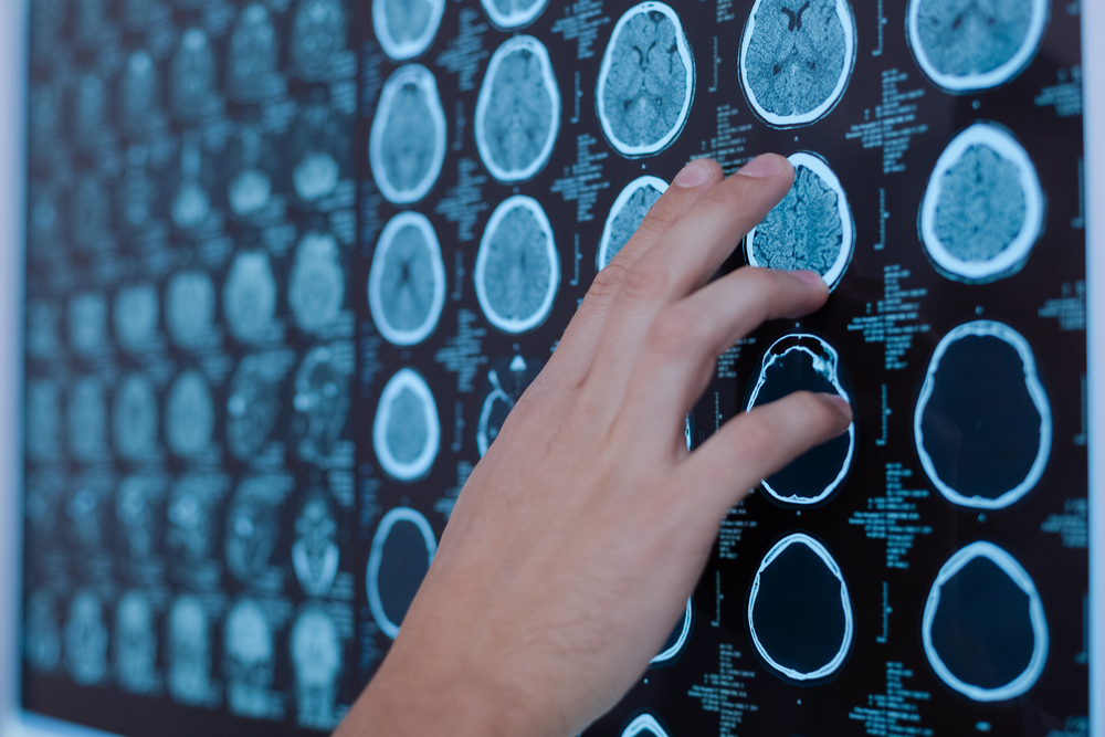
A Chinese team of researchers decided to investigate the changes in gray matter volume during cognitive degeneration by comparing Parkinson’s patients who developed mild cognitive impairment, those who did not and healthy subjects. Cognitive impairment has been linked to reduced gray matter volume.
The brain is composed of gray and white matter. The first consists of cell bodies — the control center of neurons — while the latter is made up of nerve cell projections, known as axons or fibers, connecting distinct parts of gray matter.
Ninety-four Parkinson’s patients without cognitive problems at the time of recruitment and 32 healthy subjects were included in this study. Participants underwent magnetic resonance imaging (MRI) and neuropsychological assessment at the study’s beginning and 28 months later.
Of the Parkinson’s sample, 24 subjects (16 men and eight women; mean age 63.1 years) developed disease-related mild cognitive impairment (converters) after 28 months of follow-up, while 70 individuals (43 men and 27 women; mean age 62.3 years) did not develop cognitive problems (non-converters).
Converters had significant right temporal atrophy at the beginning of the study and extensive temporal lobe degeneration 28 months later. Nonetheless, biochemical analysis showed no association between right temporal atrophy and Parkinson’s-related protein levels in cerebrospinal fluid. Sitting behind the ears, the temporal lobe is the region where sound is processed and where auditory language and speech comprehension systems are located.
Those who developed mild cognitive impairment also had progressive bilateral frontal lobe atrophy. Located directly behind the forehead, the frontal lobe carries out higher mental processes such as thinking, decision making, and planning.
Using DaT scan — an imaging technique that allows scientists to visualize the functioning of dopaminergic nerve cells — the team reported that loss of dopamine-producing neurons in the striatum (a brain region involved in motor control) of patients who progressed to mild cognitive impairment was correlated with right temporal atrophy.
The findings suggest that structural changes in the temporal and frontal lobes of Parkinson’s patients might be a biomarker for cognitive decline in the long term.


Mike Temple
Good morning. About ,6 years
go (and 2years before being diagnosed with Parkinson's) I underwent tests for " memory loss," after clinical tests and scans I was diagnosed with "frontal temporal dementia ". This turned out to be a wrong diagnosis Mike