Scientists create first visualization of tiny Parkinson’s protein clumps
Early alpha-synuclein aggregates in brain thought to trigger disease

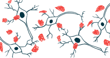
For the first time, scientists have managed to visualize tiny protein clumps in the brain that are thought to trigger Parkinson’s disease, opening the door for new insights into how the condition develops.
In a study detailing their work, the team presented an imaging method able to “generate large-scale [alpha]-synuclein aggregate maps,” or visuals of these protein clumps, in “post-mortem human brain tissue.”
Titled “Large-scale visualization of [alpha]-synuclein oligomers in Parkinson’s disease brain tissue,” the study was published in the journal Nature Biomedical Engineering.
Parkinson’s is characterized by Lewy bodies, large clumps of alpha-synuclein that are toxic to brain cells and are thought to drive the disease. It’s long been thought that alpha-synuclein first forms into small aggregates called oligomers, and then oligomers clump together to form larger Lewy bodies. Until now, however, there’s been no way to detect oligomers — which has made it difficult to study the early stages of Parkinson’s at the molecular level.
“Lewy bodies are the hallmark of Parkinson’s, but they essentially tell you where the disease has been, not where it is right now,” Steven Lee, a professor of biophysical chemistry at the University of Cambridge in the U.K. and coauthor of the study, said in a university news story describing the development of the novel imaging technique.
“If we can observe Parkinson’s at its earliest stages, that would tell us a whole lot more about how the disease develops in the brain and how we might be able to treat it,” Lee said.
Lee and a team of scientists from institutions in the U.K. and Canada figured out a way to look at alpha-synuclein oligomers using a technique called fluorescence microscopy. Very simply, this type of microscopy involves shining a laser at a sample; the sample absorbs some of the energy from the laser and emits it as light, which can then be detected to create an image.
But while this technique can detect signals from oligomers, they are very faint and are usually drowned out by background signals — sort of like how it’s impossible to see the stars during the daytime because the sun’s light drowns them out.
New technique like ‘being able to see stars in broad daylight’
In this work, the researchers essentially developed a method to reduce the background signals so that they could zero in specifically on signals from the tiny oligomers. Rebecca Andrews, a coauthor of the study who worked on the project as a postdoctoral researcher in Lee’s lab, said it’s “like being able to see stars in broad daylight.”
Using this technique — which the team dubbed Advanced Sensing of Aggregates for Parkinson’s Disease, or ASA-PD — the researchers were able to visualize alpha-synuclein oligomers in brain samples from people with and without Parkinson’s.
As expected, the scientists found that Parkinson’s patients had much higher oligomer levels. The team highlighted that oligomers were detected in healthy brains, albeit at low levels. This suggests that oligomers can form naturally, but that in Parkinson’s, something causes abnormal oligomer formation.
The researchers are hopeful that their technology can be used to further explore these mechanisms and shed light on the molecular underpinnings of Parkinson’s.
We hope that breaking through this technological barrier will allow us to understand why, where and how protein clusters form and how this changes the brain environment and leads to disease.
Sonia Gandhi, PhD, study coauthor at The Francis Crick Institute in London, noted that “the only real way to understand what is happening in human disease is to study the human brain directly.”
However, “because of the brain’s sheer complexity, this is very challenging,” said Gandhi, who co-led the research.
“We hope that breaking through this technological barrier will allow us to understand why, where and how protein clusters form and how this changes the brain environment and leads to disease,” Gandhi said.
According to Lucien Weiss, PhD, coauthor at Polytechnique Montréal in Canada who also co-led the work, this new technique could also help scientists understand other brain diseases like Alzheimer’s and Huntington’s.
“Oligomers have been the needle in the haystack, but now that we know where those needles are, it could help us target specific cell types in certain regions of the brain,” Weiss said.


