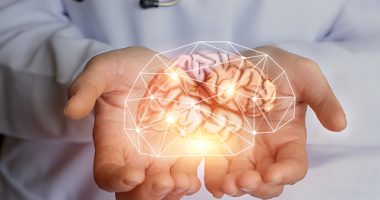
3D ‘BrainSpheres’ From Stem Cells Bring Unique Research Opportunities


Three-dimensional (3D) “BrainSpheres” made from human stem cells may be a promising new model for investigating the mechanisms that underlie Parkinson’s disease, a study has found.
When treated with certain toxins, the spheres, which are composed of functional nerve and support cells, showed molecular changes similar to those observed in commonly used animal models, the researchers reported.
“[Stem cell]-derived 3D models bring a great opportunity to study neurodegenerative diseases,” the researchers wrote, adding, “future variants of the model might be a useful tool to better understand the mechanisms of the disease and explore the use of new potential curative drugs.”
The study, “Human IPSC 3D brain model as a tool to study chemical-induced dopaminergic neuronal toxicity,” was published in Neurobiology of Disease.

In Parkinson’s, brain cells that produce a chemical called dopamine gradually degenerate, leading to the disease’s motor and non-motor symptoms.
Many animal models of Parkinson’s exist and are widely used to study the disease’s mechanisms. Usually, the models involve treatment with certain toxic compounds that mimic the dopamine cell loss observed in Parkinson’s patients.
While each has its own strengths, these existing methods may not always fully reflect the human disease, the researchers noted.
To address this, an international team of researchers developed a new technique that relies on the use of human stem cells — a type of cell with the potential to form almost any type of cell in the body — to generate a 3D “brain” in cell cultures that mimic the function of the human brain. These model brains, called BrainSpheres, contain all of the cell types, including dopamine neurons, that human brains do.
The team examined whether three toxic substances that are known to kill dopamine cells and mimic the symptoms of Parkinson’s in animal models could do the same in BrainSpheres.
Specifically, the toxic substances were: 6-OHDA, MPTP, and MPTP’s active metabolite, MPP+.
Usually, all three of the substances are toxic to living cells, or cytotoxic. In animals, they can contribute to cell death in part through mediating a process called oxidative stress. This refers to an imbalance between the production of toxic reactive oxygen species and the antioxidants that help clear them, leading to cellular stress. Disruptions in the function of mitochondria — cellular compartments involved in producing energy, are also thought to be involved.
Results overall showed that after a 24-hour exposure, all of the chemicals were able to cause cell death, lower mitochondrial function, and increase the production of reactive oxygen species as would be expected from their effects in animal models.
In parallel, the levels of several genes known to be involved in oxidative stress were increased, and levels of genes involved in mitochondria function were altered.
As is seen in animal models, the dopamine neurons seemed to be particularly susceptible to the toxic effects of the compounds.
Normally, the brain is protected from potential toxins by the blood-brain barrier, making it difficult for some substances, like 6-OHDA, to pass through. However, the researchers found that similarly to observations in animal models, the treatment could disrupt the cells of the blood-brain barrier, making it more permeable.
While all three compounds affected these processes to some degree, each seemed to have slightly different mechanisms leading to dopamine neuron loss, the researchers said. Overall, 6-OHDA was the most potent cytotoxic agent against dopamine cells in the BrainSpheres.
Overall, the findings show that BrainSpheres, when used in combination with compounds that produce Parkinson’s symptoms, produce molecular changes that are similar to those observed in animal models.
This means that the technique may be a good alternative for studying disease mechanisms, the researchers noted. Advantages of the approach include its cost-effectiveness, quickness, and potential to better translate to the human condition compared with animal models, they added.
“The use of human [stem cells] makes the model more relevant for human disease, which may be a major advantage for translational drug-discovery,” the team wrote.

The team noted that the findings are just a launch point, and that future modifications might make the technology even more useful.
For example, BrainSpheres could be produced from Parkinson’s patients with certain risk mutations or disease features, allowing for even more relevant ways of investigating the human disease.
“This approach enables studies of gene-environment interactions on a personalized level using patients [stem cells] and thus may promote precision medicine for [Parkinson’s].”