Electrical Stimulation May Correct How Brainwaves Are Affected by Parkinson’s, Study Says
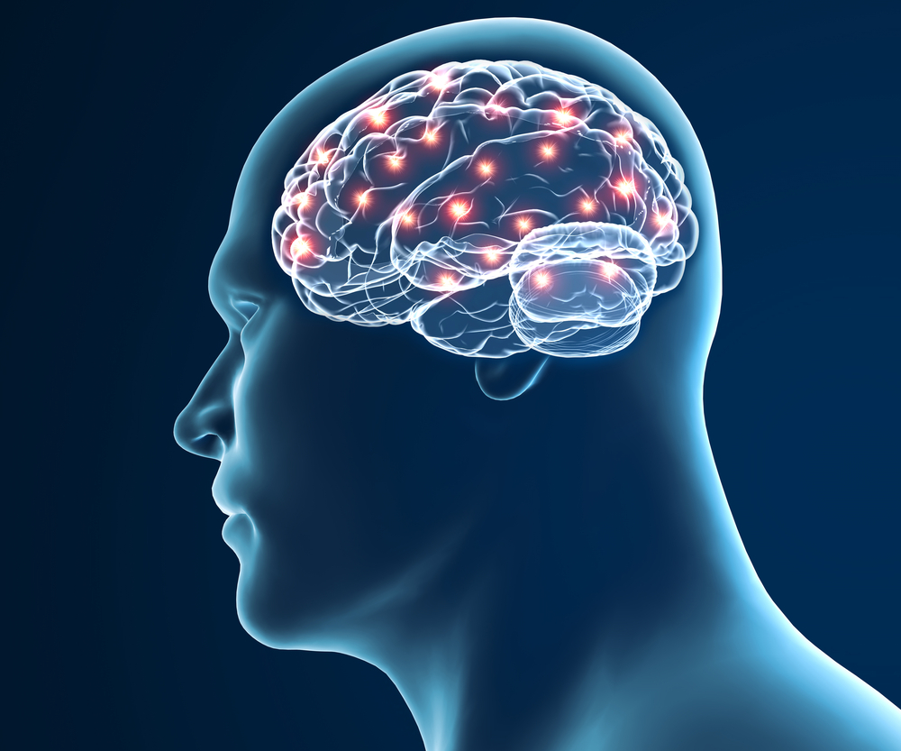


Changes in how brainwaves synchronize, known to occur in people with Parkinson’s disease, originate from distinct nerve cell networks in multiple brain regions involved in motor control, a study found.
Applying electrical stimulation to correct the abnormally coupled brainwaves may benefit these patients without the need for surgery, its researchers suggested.
The study, “Spatiotemporal features of β-γ phase-amplitude coupling in Parkinson’s disease derived from scalp EEG,” was published in the journal Brain.
Electrical activity in the brain can be measured as brainwaves (oscillations) with different frequencies. Brainwaves are produced by electrical pulses from nerve cells (neurons) communicating with each other. They are divided into different bandwidths, specifically infra-low, delta, theta, alpha, beta, and gamma, that change according to what an individual is doing and feeling.
During cognitive tasks, brainwaves of the same frequency synchronize, a process known as coupling, in specific areas of the brain.
Recent evidence has found brainwaves from different frequencies can also synchronize, a process called cross-frequency coupling. One such coupling, known as phase-amplitude coupling (PAC), is enhanced in people with Parkinson’s disease and occurs between the beta frequency bands (12–30 Hz) and gamma frequency bands (30–80 Hz).
A non-invasive EEG measuring beta-gamma PAC brainwaves can distinguish between patients and healthy individuals, as well as measure the impact of therapies that improve mobility.
However, using EEG to determine the exact location in the brain where pathological (disease-related) coupling originates has not been investigated. Furthermore, the location of coupling and its relationship to motor impairment remains unclear.
Researchers at the Leipzig University Hospital in Germany analyzed EEG recordings done in 19 people with Parkinson’s disease (13 men and six women), along with 20 age- and sex-matched healthy controls.
The team used computation techniques based on mathematical models to locate abnormal brainwave coupling within the brain, and to explore a connection with motor impairment.
Although beta-gamma phase-amplitude coupling was similar across the brain between patients and controls, a comparison of specific regions found differences.
In patients, beta-gamma phase-amplitude coupling was enhanced in the dorsolateral prefrontal cortex (executive function), premotor cortex (voluntary movements), the primary motor cortex (voluntary movements), and somatosensory cortex (processing sensation).
These brainwave coupling enhancements were also seen in the brain hemisphere opposite (contralateral) to the side of the body more affected by Parkinson’s symptoms.
Compared with controls, significant differences in beta-gamma phase-amplitude coupling were found within the same distinct brain locations, as well as between these locations. These results suggested that, in Parkinson’s patients, “abnormally enhanced coupling comprises distinct subnetworks in at least five brain regions,” the researchers wrote.
Next, to examine the relationship between phase-amplitude coupling and motor problems in patients, the team calculated the correlation between phase-amplitude coupling values in both brain hemispheres with total MDS-Unified Parkinson’s Disease Rating Scale (UPDRS) part III scores (which assess motor function) from the most affected body side (hemibody).
The overall beta-gamma phase-amplitude coupling related significantly with MDS-UPDRS III hemibody scores in the primary motor cortex, but not in the other brain regions.
While phase-amplitude coupling values from within the same brain locations did not significantly correlate with UPDRS hemibody scores in any brain region of interest, phase-amplitude coupling values between brain locations correlated with disabilities scores in the premotor, primary motor, and somatosensory cortex, “suggesting motor domain specificity.”
Finally, the team examined how the neural networks that generate abnormal coupled beta and gamma brainwaves between regions were spatially organized within the primary motor cortex.
The beta and gamma brainwave locations in both patients and controls were highly spatially correlated. Still, there was a tendency for the similarities among patients to be reduced compared to controls, indicating that “abnormally enhanced interactions became more prevalent between a few spatially distinct subnetworks in patients than in control subjects,” the researchers wrote.
“Enhanced phase-amplitude coupling in Parkinson’s disease patients originates from the coupling between distinct neural networks in several brain regions involved in motor control,” they added.
“Using external electrical or magnetic stimulation, we hope that in future it will be possible to correct the coupled electrical oscillations in Parkinson’s patients without the need for surgery,” said Joseph Classen, PhD, the study’s lead author, in a press release.
“With our mathematical modeling, we want to find out what characteristics such novel therapies would need to ensure their success. These new findings may represent an important building block in this respect,” said co-author Thomas Knösche, PhD.
As disease-related brainwave coupling was detected in a specific area of the cortex that is only partly involved in motor control, “perhaps the cognitive disorders that exist in some Parkinson’s patients have a common cause with motor disorders,” Classen added. Future studies will address this issue.