Differently-shaped Aggregates of Alpha-synuclein May Affect Onset of Parkinson’s


Scientists have discovered new aggregates of alpha synuclein of various shapes that may play a key role in the onset of Parkinson’s disease.
These new aggregates, which are larger than those described in previous studies, form in the presence of phospholipids — large fatty molecules that are the basic components of cell membranes.
According to researchers, future studies investigating the structure of these aggregates in more detail may help them distinguish those that are harmless from those associated with Parkinson’s, and lead to the development of new treatments for the disease.
Their findings were described in the study, “A series of helical α-synuclein fibril polymorphs are populated in the presence of lipid vesicles,” published in the journal Parkinson’s Disease.
One of the hallmark features of Parkinson’s is the progressive degeneration of dopamine-producing neurons located in the substantia nigra — a brain region responsible for movement control — due to the accumulation of toxic clumps of alpha-synuclein, known as Lewy bodies.
Until now, many studies have described the structure of alpha-synuclein, mostly by analyzing purified forms of the protein isolated from different sources. However, these studies have disregarded that within the brain alpha-synuclein closely interacts with phospholipids, likely participating in a series of processes that are important for neuronal function.
Now, a group of researchers from the University of Bath, U.K., have discovered that in the presence of phospholipids, alpha-synuclein fibers assemble differently, forming larger aggregates of various shapes that are different from those reported in previous studies.
In their experiments, researchers placed phospholipds and alpha-synuclein within the same test tube, and then used transmission electronic microcospy (TEM) — a high-resolution imaging technique — to visualize the structure of the aggregates that formed.
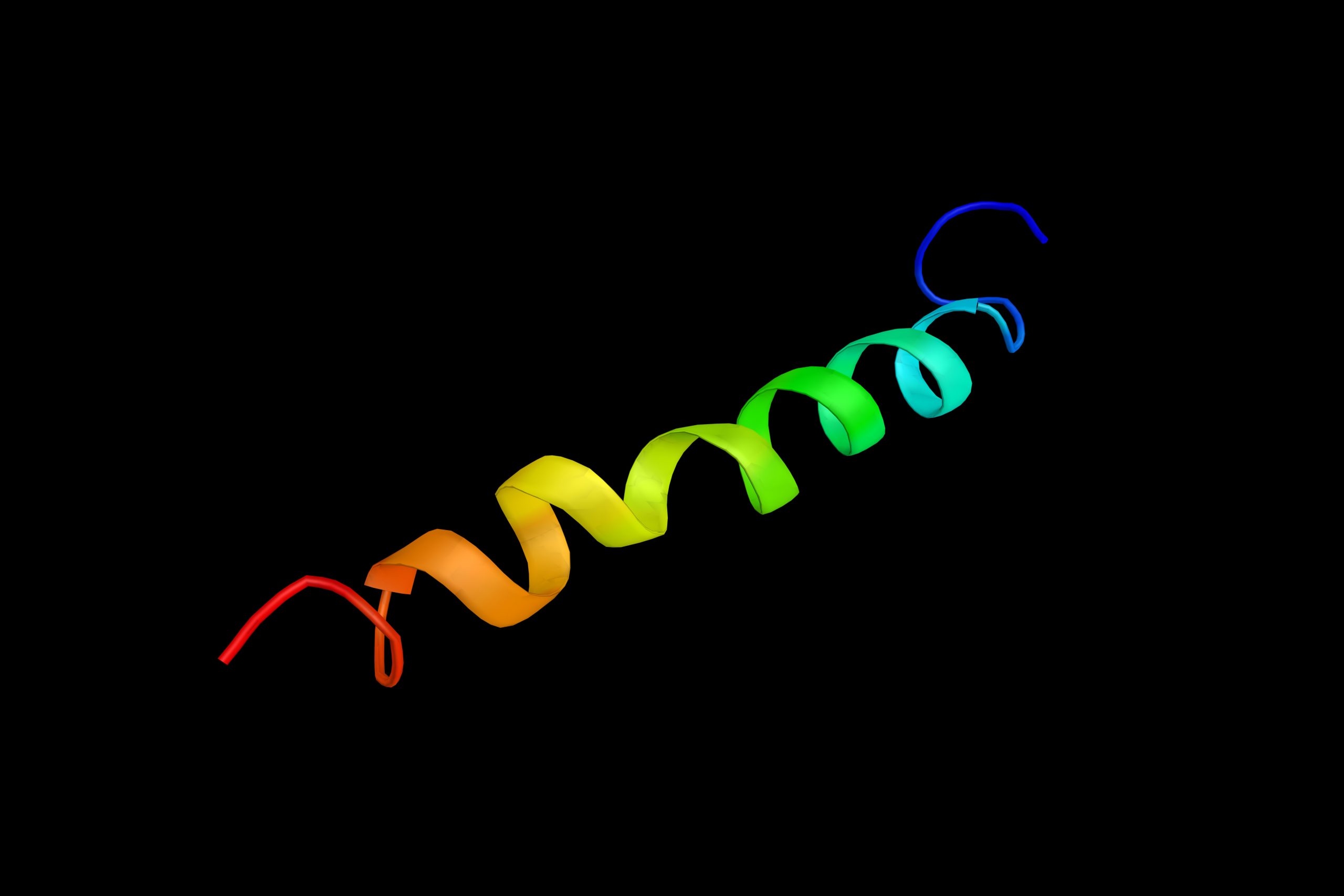
After doing so, they found the new aggregates could have different shapes, ranging from flat ribbons to wave-like structures and helices of different levels of thickness.
“We know that these misfolded proteins are heavily implicated in Parkinson’s disease,” Jody Mason, PhD, professor at the department of Biology & Biochemistry at the University of Bath, and senior author of the study, said in a press release.
“What’s more, alpha-synuclein is known to be important in neurotransmission and in cell signalling. Given its interaction with brain cell membranes, the discovery of these structures in the presence of phospholipids may have far-reaching implications in our quest to find a disease-causing form of the protein,” Mason said.
The team hopes their findings may lead to new research into the structure of alpha synuclein fibers that could shed light into which of these structures are truly involved in the onset of Parkinson’s, and which are harmless.
Once this is established, studies then may focus on testing new therapies for disease-causing forms, which will be a steppingstone in the development of a cure for Parkinson’s and other neurological conditions caused by the accumulation of toxic protein aggregates.
“These studies provide the foundation for more detailed structural analysis, and may offer new possibilities to further define disease-relevant versions of the protein that are accessible to pharmacological intervention,” the researchers wrote.
The importance of alpha-synuclein for the development of a possible cure for Parkinson’s also has been recognized by Parkinson’s UK, a charitiable organization that invested more than £3.5 million (approximately $4.6 million) to promote research in the field.
“Alpha-synuclein is known to form different structures, and researchers have become increasingly interested in which forms of the protein may be toxic, and linked to the spread and loss of brain cells in Parkinson’s — this is essential in order to develop treatments that target the right form of the protein,” said Beckie Port, PhD, research manager at Parkinson’s UK.
“By advancing our understanding of the different structures of the protein that are likely to be present inside brain cells, this University of Bath study helps pave the way for developing treatments that may one day stop the progression of Parkinson’s,” Port said.