‘Microstretcher’ Technique May Advance Parkinson’s Research


Researchers have developed a way to closely study the function of a cell’s transportation network, made up of structures called microtubules, that appears to be impaired in people with Parkinson’s disease.
This technique, called “microstretcher,” may help to better understand microtubules’ deficits and their underlying causes in Parkinson’s, as well as to identify new therapeutic targets.
“Our experimental set-up enables us to study the relationship between the deformation of microtubules and their biological functions,” Akira Kakugo, PhD, the study’s senior author at Hokkaido University, in Japan, said in a press release.
The new method was described in the study, “Regulation of Biomolecular-Motor-Driven Cargo Transport by Microtubules under Mechanical Stress,” published in the journal ACS Applied Bio Materials.
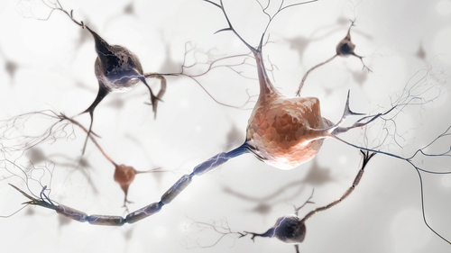
Microtubules are hollow tubular structures that provide structure not only to cells, but also form highways to transport molecules and organelles inside cells. This transport is mediated by motor proteins, namely dynein and kinesin, which move in opposite directions along microtubules’ “tracks.”
In nerve cells, microtubules are particularly involved in the transport of vesicles filled with neurotransmitters (chemical messengers used in nerve cell communication) down the axon, or nerve cell fiber, to be released as signals to other nerve cells.
Increasing evidence suggests that defective regulation of microtubules may have a role in the development of a broad range of neurodevelopmental, psychiatric, and neurodegenerative diseases, including Parkinson’s.
Microtubule dysfunction was shown to precede axon transport deficits and death of dopamine-producing nerve cells — a hallmark of Parkinson’s disease. In addition, several Parkinson’s-associated proteins, such as tau, alpha-synuclein, parkin, pink1, and LRRK2 regulate or appear to affect microtubule stability.
However, there was no experimental set-up to properly study the transport process in microtubules and the effects of disturbances in that process, until now.
Researchers at Hokkaido University and the National Institute of Information and Communications Technology, in Japan, developed a unique technique to control microtubular physical deformation and observe its effects on their transport function.
The microstretcher consists of a horizontal plate with flexible medium inside, which can be “stretched” or “compressed” using a computer system.
The team first used specific proteins to attach microtubules to the pre-stretched medium in a way that the microtubules lied parallel to the surface area and “stretching” axis. Next, dyneins bound to a fluorescent cargo were added to the microtubules.
With this system, researchers were able to evaluate the effects of stretching and compressing the flexible medium, and consequently straightening and bending the microtubules, in dynein-associated transport.
Results showed that the dynein-cargo moved faster as the microtubules began to bend, but only until the compressive strain reached about 25%, “beyond which the dynein-driven transport is retarded,” the researchers wrote.
From that point onward, the speed of dynein-associated transport started to decrease and eventually the deformation led to microtubule collapse and no dynein movement. Different speeds of motion also were observed along distinct areas of the bended microtubules.
The team noted that dynein’s faster motion in slightly bended, rather than straight, microtubules may be associated with the protein’s “walking-like” movement. Also, they believe these physical characteristics of microtubules may contribute to their functions in regulating many cellular processes.
“This work offers a technical advantage for a systematic study of the correlation between the deformation of MTs [microtubules] and its biological functions, i.e., cargo transport, as well as an opportunity to explore the interaction of deformed MTs with MT-associated proteins such as (…) tau,” the researchers wrote.
This new technique is “expected to help explain the [underlying disease-associated mechanisms] of traumatic brain injury, which mechanically stresses cells, and neurological conditions like Huntington’s and Parkinson’s diseases, in which microtubules are known to malfunction,” said Syeda Rubaiya Nasrin, the study’s first author.
Next, the team hopes to evaluate the effects of microtubule physical deformation in kinesin-driven transport along their “tracks.”
“The more we understand this process, the closer we might get to designing new nature-inspired materials that can act in a similar way,” Kakugo said.