3D Structure of Alpha-Synuclein Challenges Previous Theory of Parkinson’s


Researchers have analyzed the three-dimensional toxic form of the protein alpha-synuclein, which is directly involved in Parkinson’s disease. The findings are now challenging previous knowledge investigators had of this disease.
Parkinson’s disease is a progressive neurodegenrative disorder that belongs to the class of synucleopathies. In these type of diseases, a protein called alpha-synuclein is abnormally high. When this protein clumps together, it gives rise to small fibrils that accumulate and deposit inside brain cells, producing small inclusions called Lewy bodies.
These structures are highly toxic and often cause irreparable damage to affected nerve cells, slowly killing them. When dopamine-producing nerve cells die, patients start to experience the traditional symptoms of Parkinson’s, including lack of body balance, movement and a significant decrease in their cognitive abilities.
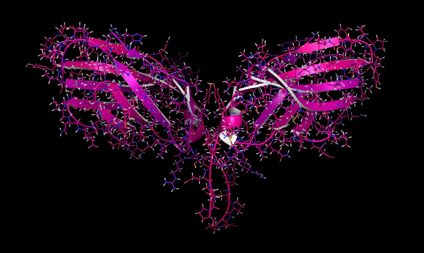
Now, in the study “Cryo-EM structure of alpha-synuclein fibrils,” which was published in eLife, Swiss researchers created an artificial form of alpha-synuclein in a lab test tube. Using cryo-electron microscopy — a high-resolution microscopy technique that uses samples frozen at very low temperatures — they were able to see for the first time the three-dimensional structure of alpha-synuclein with atomic resolution.
Discuss the latest research in the Parkinson’s News Today forum!
“Contrary to our expectations, the results seem to raise more questions than they can hope to answer,” Henning Stahlberg, the project’s principal investigator said in a press release. Stahlberg is professor of Structural Biology at the Biozentrum, University Basel, Switzerland.
Some congenital forms of Parkinson’s are caused by genetic mutations. One such mutation occurs in the SNCA gene, which encodes for alpha-synuclein. For a long time researchers suspected these mutations affected the structure of the protein, causing it to fold incorrectly and giving rise to the dangerous fibrils.
“However, our 3-D structure reveals that a mutated alpha-synuclein protein should not be able to form these type of fibrils,” said Stahlberg. “Because of their location, most of these mutations would rather hinder the formation of the fibril structure that we have found.”
In other words, if fibrils cause Parkinson’s, the genetic mutations that prevent the formation of these structures should protect against the disease. But this is not the case. So, it could be possible that a different kind of fibril or another form of alpha-synuclein are the true triggers of Parkinson’s in these patients.
More studies are necessary to understand exactly how these alpha-synuclein mutations affect the fibril structure of the protein.
In the future, investigators also hope to be able to answer other important questions about this subject, such as what is the physiological function of alpha-synuclein in the brain, and why do nerve cells die when these fibrils accumulate?