
Tau Protein Buildup in Nerve Cells Prompts Neurodegeneration
A preclinical study examined the mechanisms behind tau-mediated toxicity
Written by |
A protein “traffic jam” inside nerve cells promotes the buildup of shorter and toxic tau — a protein that forms toxic aggregates in Parkinson’s and other neurodegenerative disorders — according to a new study using fruit flies and mammalian cell lines.
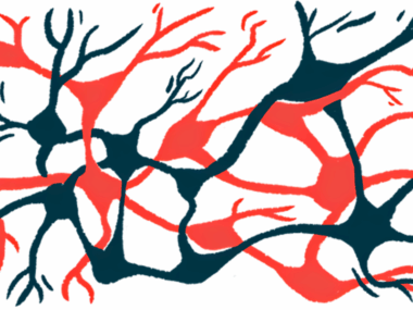
The accumulation of toxic tau resulted in fewer connections within brain nerve cells and a retraction of nerve cells’ axons, the long projections of a nerve cell that enables them to communicate.
“If we can stop or slow down the earliest disconnection of neurons, we may slow down the subsequent steps that happen as neurons start to degenerate,” said Brian McCabe, PhD, said in a press release. McCabe is the study’s lead author and the director of the Laboratory of Neural Genetics and Disease and a professor at the EPFL School of Life Sciences in Switzerland.
The study, “Retromer deficiency in Tauopathy models enhances the truncation and toxicity of Tau,” was published in the journal Nature Communications.

Parkinson’s disease is characterized by the toxic accumulation of misfolded forms of the alpha-synuclein protein within nerve cells. However, the disease also is linked with variants of the MPAT gene, which carries instructions for the tau protein. Tau can form toxic aggregates — called tau tangles — and is one of the hallmarks of Alzheimer’s, another neurodegenerative disease.
To further understand the mechanisms behind tau-mediated toxicity, researchers at the École polytechnique fédérale de Lausanne (EPFL), Switzerland, used drosophila fruit flies that were engineered genetically to express the human version of the tau protein.
Compared to normal flies who served as controls, those carrying the human tau had shorter lifespans. Prior studies had reported that increased levels of tau caused retraction and degeneration of axons.
The researchers went on to assess the impact of high tau levels in single axons. This is one of the first studies looking at neurodegeneration at the single-neuron level in an adult brain.
The analysis revealed that compared to control flies, high tau levels in a group of adult neurons, called dorsal (DC) neurons, led to a progressive loss of synaptic connections. By day 40, which corresponds to late adulthood in fruit flies, 89% of synapses were lost. This marked decreased was accompanied by the shrinkage and retraction of axons. Synapses are the junctions between two nerve cells that allows communication.
“By the time the axon was retracted, the neurons were no longer part of a functional circuit,” McCabe said. “We need to intervene in these very early stages, because when neurons are dying, the battle is already lost.”
The retromer system
The team then focused on a complex of proteins called the retromer that works as a recycling system, rescuing proteins and fats (lipids) from degradation to bring them back to the cell’s surface.
Importantly, losing the retromer complex, whose mutations are sometimes present in cases of familial Parkinson’s disease, accelerated neurodegeneration. Defects in retromer activity also has been linked with late onset Alzheimer’s, according to the researchers.
Researchers inhibited key components of the retromer system in fruit flies with high tau levels. The results showed that the inhibition led to an exacerbation of the protein’s neurotoxicity. Further experiments revealed that inhibiting the retromer activity led to a marked increase on a shorter form of the tau protein that has been linked previously with increased toxicity.
The findings were maintained when they used a mouse neuron cell line engineered to expresses the human tau protein. Lab (in vitro) studies showed that inhibiting the retromer complex resulted in an increase of a shorter tau protein.
The scientists reasoned that when retromer activity is reduced, proteins like tau linger inside the cells for longer. During this time, specialized enzymes called caspases “trim” the protein to its shorter and more toxic form.
Lessening toxicity
Inhibiting one of these particular caspases in DC neurons of fruit flies expressing the human tau protein lessened the protein toxicity, as shown by a reduction in synaptic loss and axon retraction.
Overall, these findings suggest that lowering retromer activity leads to a “traffic jam” inside cells, with tau protein lingering for longer, giving time for caspases to trim the protein into a shorter — and harmful — form of the protein.
Therapies that boost the trafficking of tau protein are thus potential candidates to lessen neurotoxicity, McCabe said. Also, if the shorter form of tau is present in human brains affected by Alzheimer’s and Parkinson’s diseases, tracking its levels could be used as a readout of the therapeutic effectiveness.
McCabe and his team will continue to use their fruit fly system to investigate the early mechanisms driving neurodegeneration.
Leave a comment
Fill in the required fields to post. Your email address will not be published.