
Sleep Problems in Parkinson’s Traced in Study to 2 Mutations That Alter How Nerve Cells Work
Written by |
Mutations in two specific genes affect how nerve cells work in the brain, disrupting sleep patterns in Parkinson’s patients, a new study suggests, and recommends a way of possibly treating this disease symptom.
The research, “ER Lipid Defects in Neuropeptidergic Neurons Impair Sleep Patterns in Parkinson’s Disease,” appeared in the journal Neuron.
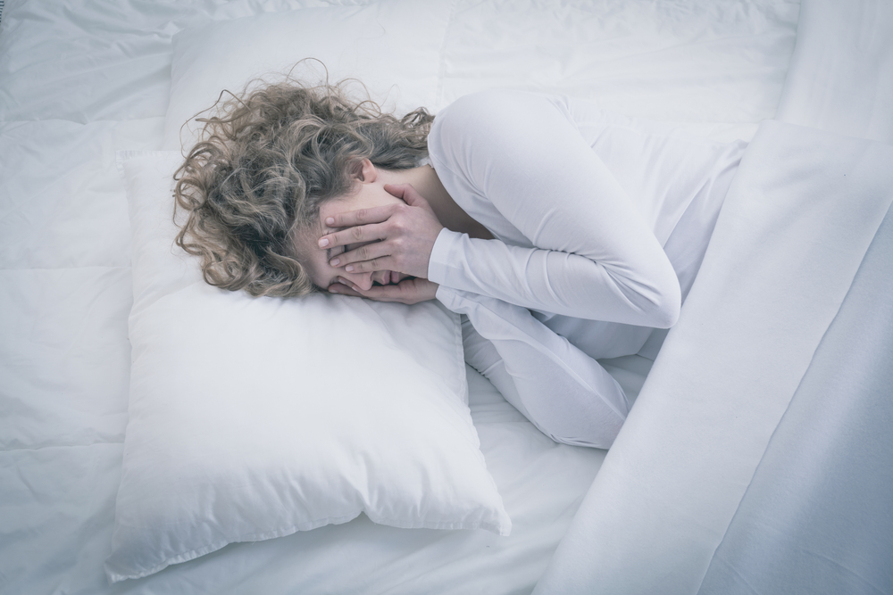
Non-motor symptoms of sporadic and familial Parkinson’s include difficulties sleeping, such as insomnia, nightmares, and restless sleep. These disturbances can occur years before Parkinson’s hallmark loss of dopamine-producing neurons and motor symptoms, and cannot be treated with dopaminergic therapy. The reasons for such disturbed sleep patterns in Parkinson’s patients, however, remains unknown.
parkin and pink1 are well-studied Parkinson’s-related genes, both broadly expressed in the brain. When they don’t function as they should, specific points of excessive contact among brain cells seem evident, the study reported, including among mitochondria — tiny cell structures that produce energy, working as the cell’s power plant. Patients with mutations in either parkin or pink1 are known to have disturbed sleep patterns.
Researchers studied these two genes in nerve cells generated from both patients’ stem cells and fruit flies, to better understand reasons for the troubled circadian — the 24-hour body rhythm – and sleep patterns in Parkinson’s patients.
Although no major defects in mitochondria or neurons were observed, results revealed that parkin and pink1 mutations led to overly abundant contact sites between mitochondria and the endoplasmic reticulum (ER) — a cellular structure particularly important in the production, folding, modification, and transport of proteins.
These contact sites caused an abnormal transfer of lipids (fats), which destroyed a component of the cell membrane (called the phosphatidylserine) with well-known implications on cognitive function and aging. The loss of phosphatidylserine, in turn, disrupted the production of vesicles that transport chemicals when nerve cells communicate and control circadian rhythms.
“Our data here suggest that understanding the basic cell biology is key,” the scientists wrote. Specifically, these alterations were found in neurons derived from a brain area called hypothalamus, which regulates sleep and wakefulness. Mutations in either gene affected the necessary increase in vesicle secretion that takes place in the brain in the hours preceding dawn, the researchers noted.
Importantly, the investigators found that feeding animals with phosphatidylserine restored the production of vesicles and eased sleep pattern disturbances.
They noted that excessive ER-mitochondrial contacts have also been reported in Alzheimer’s, amyotrophic lateral sclerosis, Huntington’s, all of which are characterized by sleep disturbances and a disrupted circadian rhythm.
However, as in Parkinson’s, little is known about sleep pattern defects in these disorders. “Our study now provides a new direction that can be tested in the context of those diseases as well,” they wrote.
And it may be of clinical relevance to patients, the researchers said, noting that their work further supports evidence that sleep impairments in Parkinson’s have a different pharmacology than its motor symptoms, which are due to dopamine loss.
“It is also important to note that the disordered circadian rhythmicity and sleep patterns are caused by neuronal dysfunction and not neurodegeneration, which implies that it can be corrected, as we show here in flies by the addition of [phosphatidylserine] to the food,” the researchers wrote.


Leave a comment
Fill in the required fields to post. Your email address will not be published.