
New Technique Based on Nanopores for the Study of Protein Structure
Written by |
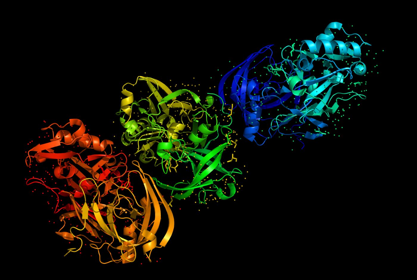
Researchers at the University of Pennsylvania recently reported a new technique based on nanopores for the study of protein structure. The technique can potentially be applied in the study of protein aggregation that occurs in neurodegenerative disorders like Parkinson’s disease. The study was published in the journal ACS Nano and is entitled “Observing Changes in the Structure and Oligomerization State of a Helical Protein Dimer Using Solid-State Nanopores”.
Parkinson’s disease is a progressive neurodegenerative disorder that develops gradually, with patients usually experiencing the first symptoms around the age of 60 or older. As the disease progresses, symptoms worsen from a barely noticeable tremor in the hands to serious difficulties in speaking, locomotion, coordination and balance. The disease is caused by the loss of the neurotransmitter dopamine due to premature death of dopaminergic neurons in the brain, which play an important role in voluntary movement and behavioral processes (mood, stress, reward, addiction). It is estimated that up to 10 million people worldwide suffer from the disease.
Disorders linked to brain cell death are often characterized by the fact that a particular protein gets misfolded (adopts an incorrect structural shape), potentially becoming toxic for nerve cells. In Parkinson’s disease, the misfolded protein that accumulates within neurons is called alpha-synuclein.
“Many researchers, (…) have observed these long tangles of aggregated peptides and proteins in diseases like Alzheimer’s and Parkinson’s, but there is an increasing body of evidence that is suggesting these tangles are occurring after the fact, that what are really causing the problem are smaller protein assemblies. Figuring out what those assemblies are and how large they are is currently really hard to do, so this may be a way of solving that problem.” explained the study’s co-author Dr. Jeffery G. Saven in a news release.
Researchers have previously developed a new method for DNA sequencing in which the DNA’s bases are read as they pass through a nanoscopic hole. Now, the team has applied this technique to proteins in order to obtain more insight into their structure.
“There are many proteins that are much smaller and harder to manipulate than a strand of DNA that we’d like to study,” noted Dr. Saven. “We’re interested in learning about the structure of a given protein, such as whether it exists as a monomer, or combined with another copy into a dimer, or an aggregate of multiple copies known as an oligomer.”
In the study, the team successfully applied their technique to a protein called GCN4-p1 leucine zipper, which has a known and common structural motif. Researchers tested different ratios of monomer and dimer versions of the protein and passed them through the nanoscopic pores. The differences in shape between the different forms of the protein resulted in a change in charge that could be detected by electronics surrounding the nanopores.
“The dimer and monomer form of the protein block a different number of ions, so we see a different drop in current when they go through the pore,” explained the study’s lead author Dr. David Niedzwiecki. “The dimer version is ‘zipped’ together, (…) It is a ‘coiled coil’ of interleaved helices that is roughly cylindrical. The monomer version is unzipped and is likely not helical; it’s probably more like a string.”
According to the authors, this technique can be applied in samples from a single individual, which can open new avenues for disease diagnosis and research. “There are no ways to amplify peptides and proteins like there are for DNA,” said the study’s senior author Dr. Marija Drndic. “If you want to study proteins from a particular source, you’re stuck with very small samples. With this method, however, you can just collect the amount of data you need and the number of proteins you want to pass through the pore and then study them one at a time as they naturally exist in the body.”
Concerning Parkinson’s disease and other disorder characterized by the aggregation of misfolded proteins, the team believes that this technique could determine whether specific proteins in a patient’s sample are aggregating or not, making it easier to obtain more insight into disease pathogenesis and evaluate its progression.
Leave a comment
Fill in the required fields to post. Your email address will not be published.