Study: Samples after death don’t accurately compare to living brains
Findings question use of postmortem brains for Parkinson's, other research
Written by |

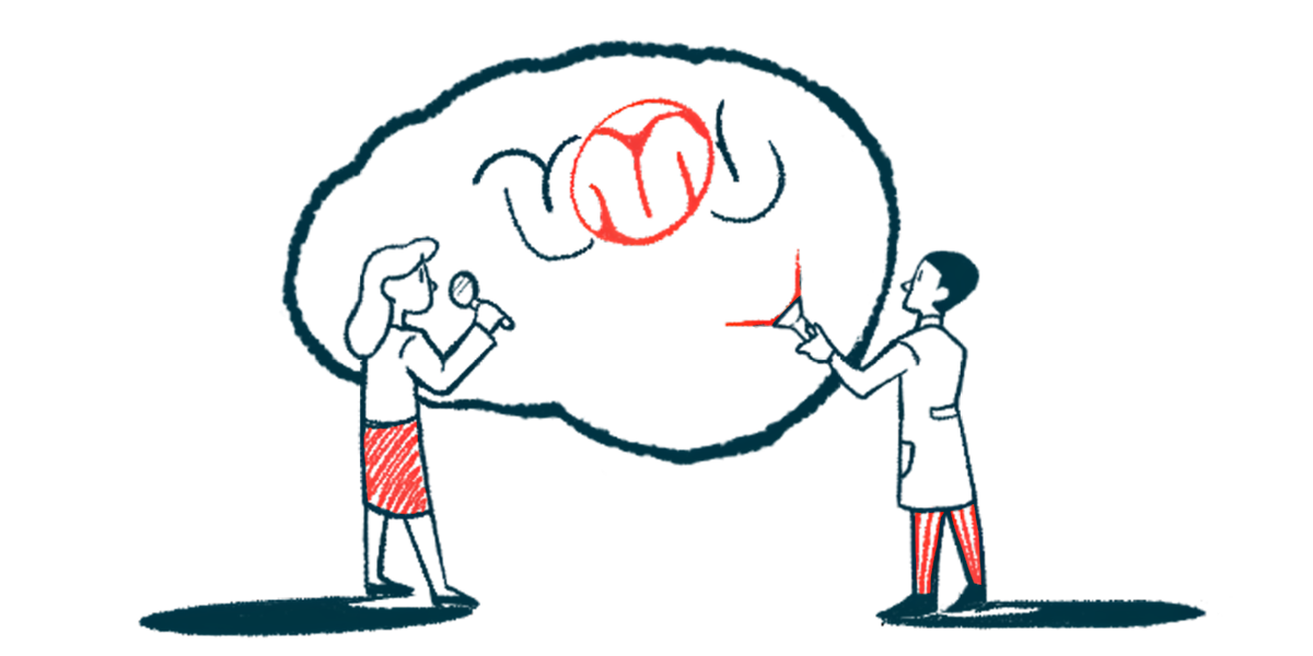
Most molecular features in the human brain differ between samples obtained from living individuals and those collected after death, according to new findings from the Living Brain Project, a multiscale investigation from The Friedman Brain Institute in New York.
This study, led by researchers at the Icahn School of Medicine at Mount Sinai in collaboration with Bpgbio, a clinical stage biopharma, shows that 95% of RNA molecules and 61% of proteins are significantly different in living and postmortem samples.
These findings challenge the longstanding view that postmortem brain samples serve as an accurate representation of the living brain, and can be used to study the molecular mechanisms —including genes, proteins, chemicals, and cell types — of neurodegenerative conditions such as Parkinson’s disease.
“Neurodegenerative diseases have not seen meaningful therapeutic progress in decades, and findings from this project suggest that we’ve been handicapped by not having access to tissue reflective of brain function,” Alexander Charney, MD, PhD, colead author and vice chair of the artificial intelligence and human health department at Icahn Mount Sinai, said in a press release from Bpgbio.
The study, “A study of RNA splicing and protein expression in the living human brain,” was published in the journal PLOS One.
Researchers at Icahn will now continue to work with Bpgbio — which leverages artificial intelligence (AI) to make sense of massive, complex data sets — to generate new insights from living brain samples and potentially develop new drugs for Parkinson’s and other neurodegenerative diseases.
“The Living Brain Project is a game changer and represents a breakthrough path forward for the field,” Charney said. “By working with Bpgbio, whose capabilities in integrating [complex] data and AI are second to none, we are now able to generate actionable insights from living brain tissue that can reshape the future of drug development in Alzheimer’s, Parkinson’s, and other devastating diseases.”
Parkinson’s brain samples collected from patients undergoing DBS surgery
Because researchers cannot collect brain samples from a living person, postmortem brain samples have been widely used as a surrogate, helping to better understand the molecular mechanisms of neurodegenerative diseases and identify promising therapeutic targets.
However, postmortem brain samples are typically collected 12-24 hours after death, and there haven’t been many robust studies supporting the assumption that postmortem brain samples are an accurate molecular representation of the living brain.
The researchers noted that the Living Brain Project was “designed to rigorously compare living and postmortem human brain samples at multiple molecular levels.”
For this study, living tissue was collected from the prefrontal cortex — a part of the brain located behind the forehead that’s responsible for attention, emotions, self-control, and decision-making — of people with neurological or neuropsychiatric conditions undergoing deep brain stimulation (DBS) surgery.
DBS, often used in Parkinson’s to help ease patients’ symptoms, is a procedure in which electrodes are placed into specific areas of the brain to modulate neural activity.
By directly accessing and characterizing living brain tissue at multiple molecular levels, we are not just challenging old assumptions — we are opening entirely new avenues for target discovery and therapeutic innovation.
To date, researchers have analyzed more than 500 living brain samples and matched postmortem controls.
“By directly accessing and characterizing living brain tissue at multiple molecular levels, we are not just challenging old assumptions — we are opening entirely new avenues for target discovery and therapeutic innovation,” said Michael A. Kiebish, PhD, Bpgbio’s vice president of platform and translational sciences.
Kiebish said “these insights will fuel” the company’s AI platform and “accelerate the development of the next generation of treatments for neurodegenerative disease.”
Living Brain Project setting up ‘new frontier in neuroscience research’
For the current study, the scientists studied 288 brain samples obtained from 171 living participants and compared them with 246 postmortem control samples from three brain banks. Most samples were collected from men and individuals with Parkinson’s. Living and postmortem samples were matched as much as possible for age, sex, and the size of tissue collected.
Results from the study demonstrated that 74% of primary RNA molecules and 70% of mature messenger RNA (mRNA) molecules — the intermediate molecule derived from DNA that guides protein production — and 61% of the proteins differed between living and postmortem brain samples.
Primary RNA transcripts are unprocessed RNA molecules that contain both coding and noncoding regions of a gene. These molecules are then modified via a process called splicing to keep only the coding regions, forming the mature mRNA molecules that are used for protein synthesis.
Researchers also observed significant differences in the splicing of 73% of primary RNA transcripts, which can affect the final protein product. The pattern of RNA-protein correlations was also markedly different between living and postmortem tissue.
Niven R. Narain, PhD, president and CEO of Bpgbio, called teaming up with Charney and the Mount Sinai investigators “a testimony to the meaning of true collaboration and … [the partners’] shared commitment to breaking new ground in medicine.”
The goal, Narain said, is new treatments based on these discoveries.
“The Living Brain Project paves the way for a new frontier in neuroscience research, and it has the potential to transform how future medicines will be developed,” Narain said. “We look forward to collaborating with Mt. Sinai and commercial partners who share our vision to accelerate novel insights to create impact for patients and families.”


