
Link Seen Between 2 Key Features of Early-onset Parkinson’s and PARK2 Gene
Written by |
A direct causal link was found between two major underlying features of Parkinson’s disease — mitochondrial defects and lysosomal malfunction — in dopamine producing neurons that lacked the PARK2 gene, which is mutated in some cases of early-onset disease, a study discovered.
Further research is needed to determine if these findings can lead to treatment approaches that address such defects.
The study, “Lysosomal perturbations in human dopaminergic neurons derived from induced pluripotent stem cells with PARK2 mutation,” was published in Nature Scientific Reports.
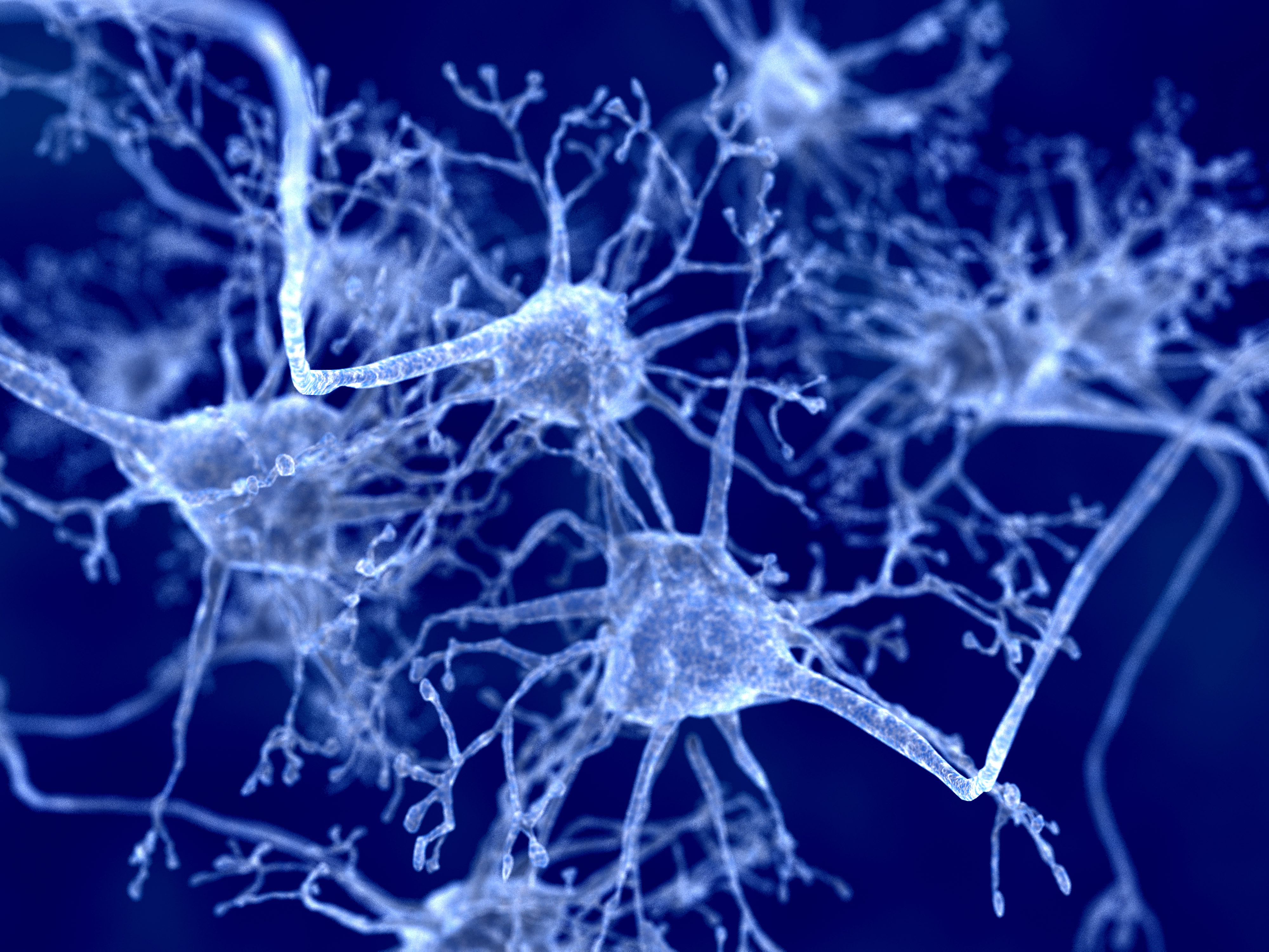
In Parkinson’s disease, nerve cells — known as dopaminergic neurons — gradually begin to malfunction or die in a region of the brain called the substantia nigra. These cells typically produce the neurotransmitter dopamine, which passes signals between neurons responsible for movement and coordination.
People with early-onset Parkinson’s disease are more likely to carry mutations in specific genes associated with the disease.
Mutations in the PARK2 gene (or PRKN), which carries the instructions for a protein called parkin, have been identified as the most common cause of early-onset Parkinson’s.
Parkin is an enzyme that chemically tags damaged and excess proteins that are part of small structures within the cell (organelles) known as mitochondria — the energy-producing centers in cells.
Tagged proteins are then transported to another organelle called the lysosome, which degrades these proteins so they can be recycled. This system acts as a quality control system for cells by disposing of damaged, deformed, and excess proteins.
However, neither the exact relationship between mitochondria and lysosomes in PARK2-related Parkinson’s disease, nor their role in disease progression are well defined.
Scientists at the University of Southern Denmark created dopaminergic neurons with and without a functioning PARK2 to mimic the cellular environment found in early-onset Parkinson’s. The neurons were then analyzed and compared to identify disease-related molecular mechanisms.
The dopaminergic neurons were created from a type of stem cell called induced pluripotent stem cells, which are generated directly from adult cells and are then be reprogrammed to become any cell of choice.
Neurons without PARK2 — called PARK2 KO — grew into midbrain dopaminergic neurons with the same efficiency as the normal (control) neurons.
An analysis found two proteins associated with lysosomes (LAMP1 and LAMP2A) at significantly higher levels in PARK2 KO neurons than in control neurons, indicating increased lysosomal content. Microscopic imaging showed significantly changes in the shape of the altered lysosomes; they appeared to be larger as well as more abundant.
Overall, lysosome activity was found to be more than 30% lower in PARK2 KO neurons, and the activity of two key lysosomal enzymes was reduced as well. The alterations in lysosome activity were seen to affect the protein recycling process directly.
Interestingly, the activity of a lysosomal enzyme called GCase, which is encoded by the GBA gene and linked to both Parkinson’s disease and a lysosomal storage disorder called Gaucher disease, was significantly increased in PARK2 KO neurons.
According to the scientists, this “points to the activation of compensatory mechanisms to increase GCase activity” and may reflect a direct connection between PARK2 and GBA.
Alterations in the mitochondria of PARK2 KO neurons were found as well. These mitochondria were often accumulated in the area just outside the cell nucleus, and they appeared swollen with irregular internal shapes. In contrast, mitochondria in the control cells were more evenly distributed around the cell, with a typical oval appearance and well organized internal structure.
Treating the control neurons with a chemical that causes mitochondrial stress led to an increase in lysosomes to a level similar to that of untreated PARK2 KO neurons, “strongly indicating that mitochondrial function impairment leads to lysosomal accumulation.”
Significantly higher production of reactive oxygen species (ROS), which damages mitochondria, were detected in PARK2 KO neurons. Treating cells with an antioxidant that blocks ROS production was able to lessen oxidative stress and significantly decrease the mitochondrial and lysosomal areas when compared with untreated neurons.
Of note, oxidative stress is an imbalance between the production of ROS and the ability of cells to detoxify them, leading to cellular damage and death.
“These data provide initial evidence of the possible beneficial effects of antioxidant treatment on both mitochondria and lysosomes, however, further research is required for accurate understanding,” the researchers wrote.
“Our results indicate that the loss of parkin causes several lysosomal perturbations affecting their abundance, morphology, content, and activity,” they concluded. “Taken together … these findings indicate a causal link between two major pathological features of [Parkinson’s disease], namely mitochondrial defects and lysosomal dysregulation.”
“Future efforts should focus on the identifying the molecular mechanistic pathways that connect mitochondrial and lysosomal perturbations with the aim of developing therapeutic approaches for [Parkinson’s disease],” they added.


Leave a comment
Fill in the required fields to post. Your email address will not be published.