Brain MRIs Can Be Used to Detect Early Signs of Parkinson’s Cognitive Impairment, Study Suggests

Ana Pena, PhD avatar

by Ana Pena, PhD |

Share this article:

Share article via email
brain MRI

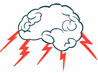
Brain magnetic resonance imaging (MRI) scans could be used to detect early and subtle markers of cognitive impairment in people with Parkinson’s, which may help predict patients’ prognoses and disease progression, a recent study suggests.

Such early detection also allows people with the neurodegenerative disease to start appropriate care strategies earlier, the researchers say.

The results of the study, “Texture features of magnetic resonance images: A marker of slight cognitive deficits in Parkinson’s disease,” were published in the journal Movement Disorders.

Cognitive impairment is a common non-motor symptom of Parkinson’s disease and a cause of significant disability for patients and a burden for caregivers. The extent and progression of cognitive deficits vary, with about 20.3% to 60.5% of individuals experiencing mild cognitive impairments (MCI). In more severe cases, such impairment can result in dementia.
Those at-risk can benefit from early detection of cognitive alterations, which allows them to initiate appropriate care strategies such as cognitive stimulation therapy. Such therapy can result in a marked improvement in cognition and quality of life, according to researchers. However, standard neuropsychological assessments are time-consuming and not easy to do in routine clinical practice. Moreover, such evaluations can be influenced by medication, pain, anxiety, and other factors.
Therefore, additional markers of cognitive deficits are needed for Parkinson’s patients, the researchers say. One potential option is the use of magnetic resonance imaging (MRI), an imaging exam that uses a powerful magnetic field, radio waves, and a computer to produce detailed pictures of the body’s internal structures.
“Texture features” — a well-known method of MRI image processing used for medical purposes — could offer insights on subtle brain changes.  These features could be used to detect the damage to brain cells, long before any symptoms of cognitive impairment develop.
Recognizing the potential of this method, a team of French researchers now tested whether such signals could be used as early markers of Parkinson’s cognitive impairments — “potentially even before the atrophy [loss of brain volume, a usual sign of cognitive decline] becomes manifest,” they said.

The team investigated if MRI texture analysis is sensitive enough to be an early marker of cognitive alterations, specifically of cognitive slowing, in Parkinson’s patients.

They analyzed brain MRI scans of 102 people with Parkinson’s from centers in Lille, France, and Maastricht, the Netherlands, who were involved in a previous study.

Based on tests of attention, memory, executive function, language, and visuospatial functions, three groups of patients were considered for the study. These groups were cognitively intact patients (PDCN); cognitively intact patients with slight cognitive slowing (PDCN-S); and patients with mild cognitive deficits, particularly in executive functioning (PD-EXE).

A group of 17 age‐matched healthy people (controls) was included for comparison. All participants were examined on a 3T whole-body scanner and T1‐weighted images were acquired.

Six regions of the brain previously reported to suffer from atrophy (volume loss) in Parkinson’s patients with cognitive impairments were specifically chosen by the researchers for analysis. These regions were the thalamus, the hippocampus, the puramen, the pallidum, the caudate nucleus, and the amygdala.

The researchers found that values for two texture features — skewness and entropy — could distinguish individuals who had normal cognition from those with slight cognitive slowing, and from those with mild impairments. Skewness is a parameter that quantifies the asymmetry of the intensity of MRI signals. Entropy represents the degree of uncertainty of the texture intensity.

These texture features were at three specific regions in the brain: the hippocampus, the thalamus, and the amygdala.

The values for these features gradually decreased in those patients with worse cognitive function, suggesting it is possible to detect early cognition deficits in people with Parkinson’s using MRIs. The researchers noted that the best performances regarding sensitivity and specificity were obtained by measuring skewness in the hippocampus. In fact, skewness in the hippocampus was a significant marker of slight cognitive slowing.
“Our results suggest that hippocampal neurons could be affected very early in PD patients, even before atrophy can be detected with commonly used methods, and this could cause a general slowing of information processing,” the researchers said.
“These results support the assumption that signal alterations associated with Parkinson’s disease–related cognitive decline can be captured very early by texture analysis,” they added.
The researchers believe that brain MRI imaging could be combined with other methods, such as cognitive assessments and electroencephalograms. That would allow scientists to build a combined model “not only for the profiling but also for the prognosis and the prediction of evolution” of cognitive impairment, they said.