Blocking 2 proteins could slow Parkinson’s progression: Study
Protein pair found on brain neurons may be key to disease spread
Written by |
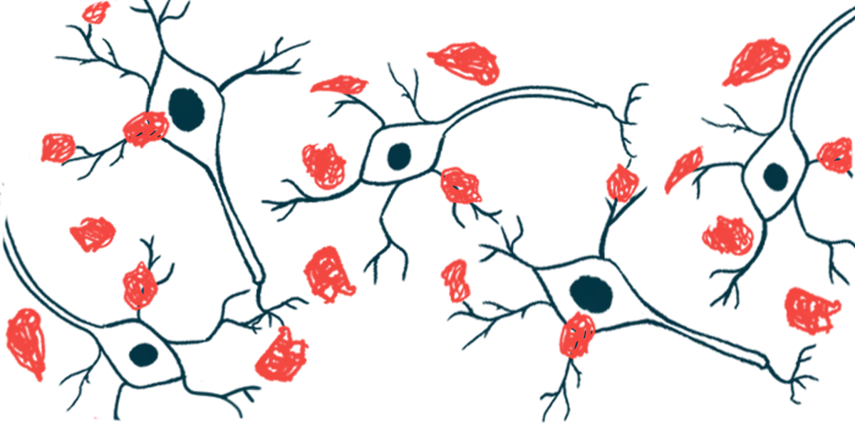
- Parkinson's progression is linked to toxic alpha-synuclein clumps spreading between neurons.
- Two proteins on neurons facilitate this spread and neuronal death.
- Blocking these proteins could slow Parkinson's progression and ease symptoms.
Two proteins found on the surface of dopaminergic neurons, the nerve cells that are lost in Parkinson’s disease, may play a crucial role in the spread of toxic alpha-synuclein clumps throughout the brain, and blocking them could slow or halt disease progression.
That’s according to a study from Yale School of Medicine, which found that alpha-synuclein transmission to neighboring neurons was reduced in cellular and mouse models lacking functional versions of the proteins, mGluR4 and NPDC1. In mice, the absence of these proteins also lessened Parkinson’s-like symptoms and improved survival.
“We have an aging population. How we can stop or slow neurons from dying is an enormous problem,” Stephen Strittmatter, MD, PhD, professor at Yale School of Medicine and senior author of the study, said in a university news story. “This is really the time to make some inroads into figuring out how to slow it down.”
The study, “mGluR4–NPDC1 complex mediates α-synuclein fibril-induced neurodegeneration,” was published in Nature Communications.

How does disease spread?
Parkinson’s disease is caused by the progressive dysfunction and loss of dopaminergic neurons, the nerve cells that produce the signaling molecule dopamine, in a brain region called the substantia nigra. The loss of dopamine signaling leads to the disease’s motor symptoms, such as tremors, slowed movements, and balance and gait issues.
The accumulation of toxic clumps of misfolded alpha-synuclein protein and their spread throughout the brain is believed to be a driver of neuronal loss in Parkinson’s disease. However, the mechanisms by which alpha-synuclein clumps spread among neurons remain unknown.
“If we understood how [alpha-synuclein] gets into neurons, we could perhaps block or slow down the progression of the disease,” Strittmatter said. To do that, he said, “we need to understand the molecular mechanism of how it spreads.”
This study suggests that mGluR4 and NPDC1 may be essential in this process.
The researchers screened more than 4,000 membrane proteins to identify those that bind to toxic clumps (fibrils) of the alpha-synuclein protein. While they identified 16 candidates, they focused on mGluR4 and NPDC1 because these proteins are specifically found on the surface of dopaminergic neurons in the substantia nigra.
To test whether these two proteins could help alpha-synuclein spread to other neurons, the scientists used a mouse model lacking functional mGluR4, injecting alpha-synuclein fibrils into the animals’ striatum, a brain region involved in motor control that is also affected in Parkinson’s disease.
In both groups, the injection caused alpha-synuclein to clump in the brain. However, while the normal mice suffered a significant loss of dopamine neurons and developed motor deficits, the mice lacking mGluR4 were protected from neuron death and maintained their motor function. This indicates that “mGluR4 is required for cell loss induced by [alpha-synuclein clumping],” the researchers wrote.
Similar results were observed in mice without functional NPDC1. In another mouse model expressing human mutant alpha-synuclein, which presents with neuronal loss, progressive motor deficits, and shorter survival, the absence of mGLUR4 or NPDC1 prevented some of these effects.
Further experiments in cells demonstrated that the two proteins form a physical complex that acts as a gatekeeper, allowing the toxic clumps to enter and kill healthy neurons. Thus, researchers investigated whether eliminating both proteins could have a cumulative effect.
In neurons derived from mice missing both proteins, there was a more significant reduction in the binding of alpha-synuclein clumps compared to neurons from mice missing only one of the two proteins.
The researchers said the results support “a critical role for the mGluR4 – NPDC1 physical complex in neuronal binding of [alpha-synuclein fibrils].”