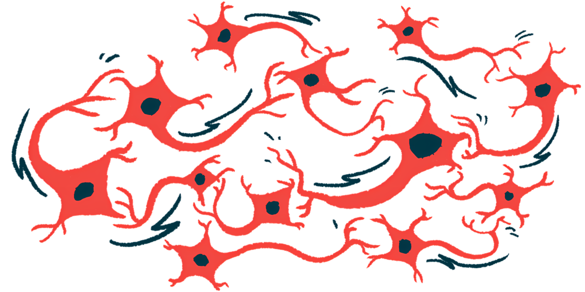
Aged neurons don’t respond normally to stress: Study
They're more apt to contribute to neurodegenerative diseases like Parkinson's
Written by |
Aging interferes with the way neurons, or nerve cells, respond to stress, making them more susceptible to damage and more likely to contribute to neurodegenerative diseases such as Parkinson’s, according to a study in cells and brain tissue.
“It’s the neuronal equivalent of being so stressed that you catch a cold,” Kevin Rhine, PhD, the study’s first author and a postdoctoral research fellow at University of California (UC) San Diego’s Sanford Stem Cell Innovation Center, said in a university news story.
The study, “Neuronal aging causes mislocalization of splicing proteins and unchecked cellular stress,” was published in Nature Neuroscience.

Aged neurons appear to forget about stress response
As people grow older, their risk of developing Parkinson’s and other neurodegenerative diseases increases. Such diseases drive “a substantial portion of the public health burden,” the researchers wrote. However, what exactly happens inside aged neurons remains unclear.
“Aging has been a black box for a long time,” said Gene Yeo, PhD, who led the study. “Nobody is really sure what an aged neuron looks like, how it behaves, or how it’s different from a young neuron.”
Yeo is director of the Sanford Stem Cell Innovation Center and a professor at UC San Diego School of Medicine.
To learn more, Yeo’s team collected skin cells from healthy elderly people and reprogrammed them into neurons while maintaining any signs of aging. The researchers then compared these lab-grown aged neurons to brain tissue from both humans and mice.
In these aged neurons, the activity of more than 4,000 genes changed. Specifically, while genes for RNA-binding proteins, which help process the way cells edit genetic instructions before using them, were still present, the actual helper proteins were found at lower levels.
Many of these proteins were no longer in the cell’s nucleus, where they normally work, and had moved to the cytoplasm, the fluid part of the cell outside its nucleus.
“We think that aged neurons are prioritizing other proteins and forgetting about the stress response and about RNA-binding proteins that keep everything running smoothly,” Yeo said. Without enough of these proteins, neurons can’t properly edit genetic instructions.

Key protein missing from stress granules
One protein researchers found to be behaving unusually is called TDP-43. Like some other proteins linked to neurodegenerative diseases, TDP-43 can form toxic aggregates that build up in or around neurons, causing damage. Although the buildup of TDP-43 is commonly found in people with amyotrophic lateral sclerosis (ALS), its accumulation has been found in Parkinson’s.
Normally, when cells are stressed, the certain proteins move into stress granules — temporary storage areas in the cell that form during stress. However, in aged neurons, these proteins didn’t move into the granules as expected. TDP-43 was also missing from these granules, which might explain why it tends to clump together.
Aging is one of the primary risk factors for neurodegeneration, and our study demonstrates that aging alone mechanistically and functionally destabilizes RNA metabolism [chemical processes] in neurons.
Old neurons may be already under constant stress, which interferes with their normal response to stress. This constant stress also affects other processes, such as ubiquitylation, which helps remove damaged proteins or other material no longer needed by cells.
To confirm that their lab-grown aged neurons reflect real aging in the brain, the researchers studied brain tissue from people ages 30 to 50 and 80 to 90. In older brain tissue, RNA-related pathways — especially RNA-binding proteins — were reduced. This matched what they had seen in the aged neurons.
In older brain tissue, TDP-43 was more often mislocated in the cytoplasm, just like in the aged neurons. Older brain tissue also showed signs of chronic (long-lasting) stress, suggesting aged neurons may accurately reflect key features of aging in the human brain.
“Aging is one of the primary risk factors for neurodegeneration, and our study demonstrates that aging alone mechanistically and functionally destabilizes RNA metabolism [chemical processes] in neurons,” the researchers wrote.