3D Structure of Brain Alpha-Synuclein More Heterogeneous Than Previously Thought, New Study Reports
Written by |

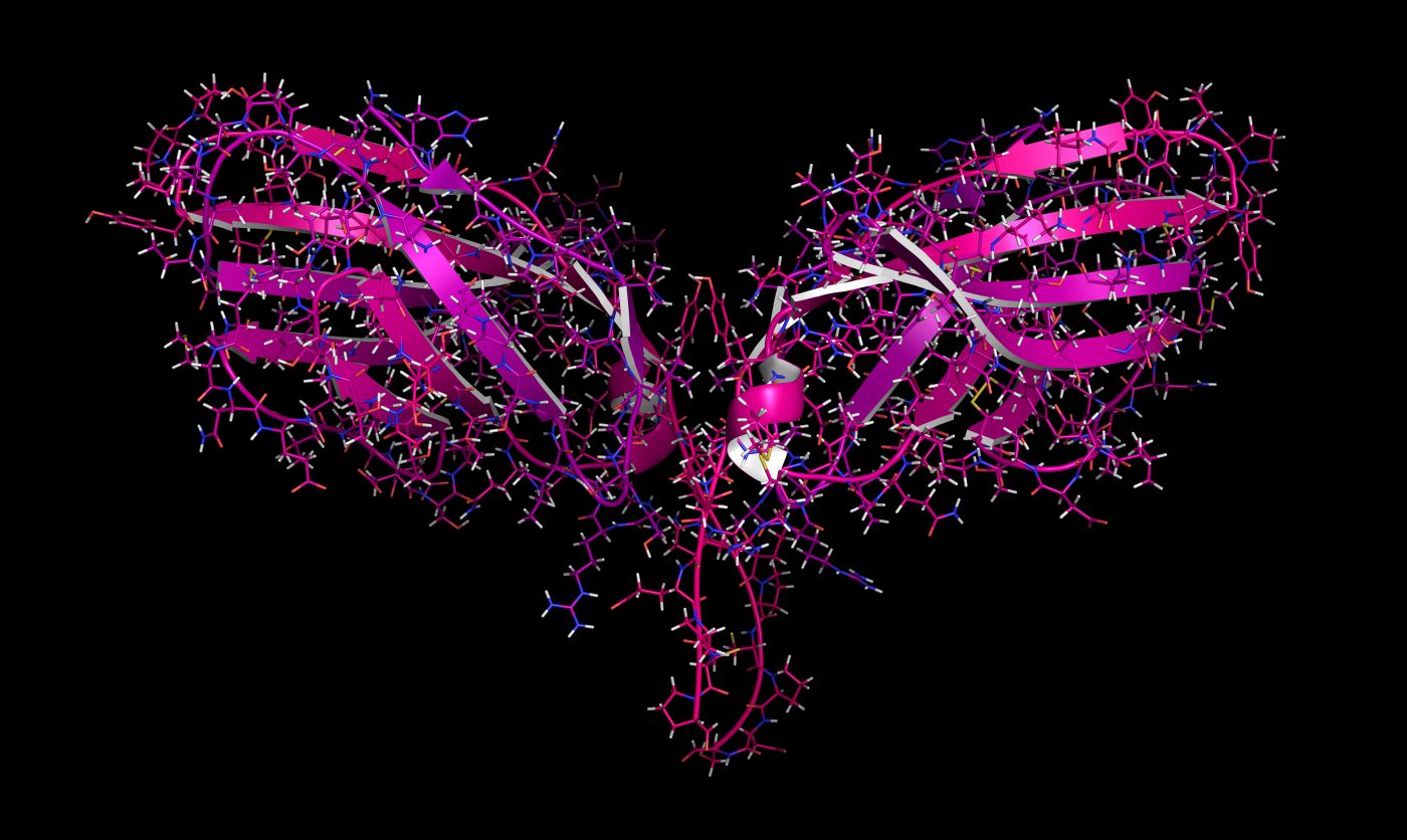
Clumps, or aggregates of alpha-synuclein protein in the brain, a hallmark of Parkinon’s disease, are more heterogeneous than previously thought, according to a new study. Moreover, the protein’s 3D-structure is different in Parkinson’s patients as compared with lab-made versions.
These findings may hold implications for the development of treatments targeting alpha-synuclein, as new therapies need to take into account what kind of shape the protein adopts within nerve cell aggregates.
The study, “Structural heterogeneity of α-synuclein fibrils amplified from patient brain extracts,” was published in the journal Nature Communications.
Parkinson’s disease and multisystem atrophy (MSA) belong to a class of neurodegenerative disorders called synucleinopathies that are characterized by the accumulation of misfolded alpha-synuclein proteins.
These abnormal protein aggregates are toxic, and mainly accumulate in Parkinson’s disease in dopamine-producing nerve cells — those responsible for releasing the neurotransmitter dopamine. Dopamine is a chemical messenger that allows nerve cells to communicate and, among other functions, helps regulate movement.
“These deposits successively appear in various areas of the brain. They are a disease hallmark,” Markus Zweckstetter, a professor and research leader at the German Center for Neurodegenerative Diseases (DZNE) and the Max Planck Institute for Biophysical Chemistry (MPI-BPC), said in a press release.
“There is evidence that these aggregates are harmful to neurons and promote disease progression,” he added.
Given the key role of alpha-synuclein aggregates in the development and progression of Parkinson’s disease and other neurodegenerative disorders, these protein clumps represent potential therapeutic targets. Treatments could either prevent the aggregates from forming or eventually clear them from within the brain.
To identify which specific sites on alpha-synuclein can be targeted by potential therapies, researchers need to have an understanding of the complex structure these proteins acquire once they form aggregates. However, current knowledge is limited to aggregates that are established in the lab, in a test tube, which may be different from what actually happens in patient’s brains.
“Previous studies investigated the molecular structure of aggregates that were synthesized in a test tube. We asked ourselves how well such artificially produced specimens reflect the patient’s situation. That is why we studied aggregates generated from tissue samples from patients,” Zweckstetter said.
Zweckstetter and his team collaborated with researchers in Australia and South Korea to analyze the structure of alpha-synuclein aggregates derived from brain samples of individuals with Parkinson’s disease (five patients) and MSA (five patients). They then compared them to those artificially produced in the lab.
The brain samples were taken from a region of the brain called the amygdala, which plays a key role in regulating emotions and memory, and is known to be affected in Parkinson’s.
The brain-derived alpha-synuclein aggregates were structurally different to those generated in the lab, the researchers found.
Alpha-synuclein proteins found within aggregates contained a structure known as “beta sheets”— whose orientation was of relevance — and their molecular backbone was twisted in a way that the protein was mainly two-dimensional. That is in contrast with a three-dimensional structure.
Within aggregates, alpha-synuclein proteins stick together in layers. However, this folding was not seen across the whole protein, with certain areas remaining unstructured.
The scientists also found that alpha-synuclein aggregates from Parkinson’s patients showed more diverse structures than those from people with MSA. That may reflect “the greater variability of disease phenotypes [manifestations] evident in PD [Parkinson’s disease]”, the researchers said.
“Proteins of MSA [multisystem atrophy] patients differed from those of Parkinson’s patients. If one looks at the data more closely, you notice that the proteins of the MSA patients all had a largely similar shape. The proteins of the patients with Parkinson’s were more heterogeneous. When comparing the proteins of individual Parkinson’s patients, there is a certain structural diversity,” said Timo Strohäker, first author of the study.
The researchers noted that “although larger patient numbers are required to link [alpha-synuclein] aggregate structure with symptomatic heterogeneity of the disease, our data point to the possibility that [alpha-synuclein] aggregate structure could be specific for individual patients or certain subtypes of [Parkinson’s disease].”
The findings of differently structured alpha-synuclein proteins among different Parkinson’s patients contrasts with current hypotheses theorizing that each neurodegenerative disease is associated with a single, defined structure of alpha-synuclein protein.
“The variability of Parkinson’s disease could be related to differences in the folding of aggregated alpha-synuclein,” Zweckstetter said. “This would be in contrast to the ‘one disease-one strain’ hypothesis, that is to say that Parkinson’s disease is associated with one, clearly defined aggregate form.”
“However, in view of our relatively small sample of five patients, this is just a guess,” he added. “Yet, our results certainly prove that studies with tissue samples from patients are necessary to complement lab experiments in a sensible way.”


