Supporting Brain Cells, Astrocytes Also May Contribute to Parkinson’s Disease, Study Suggests
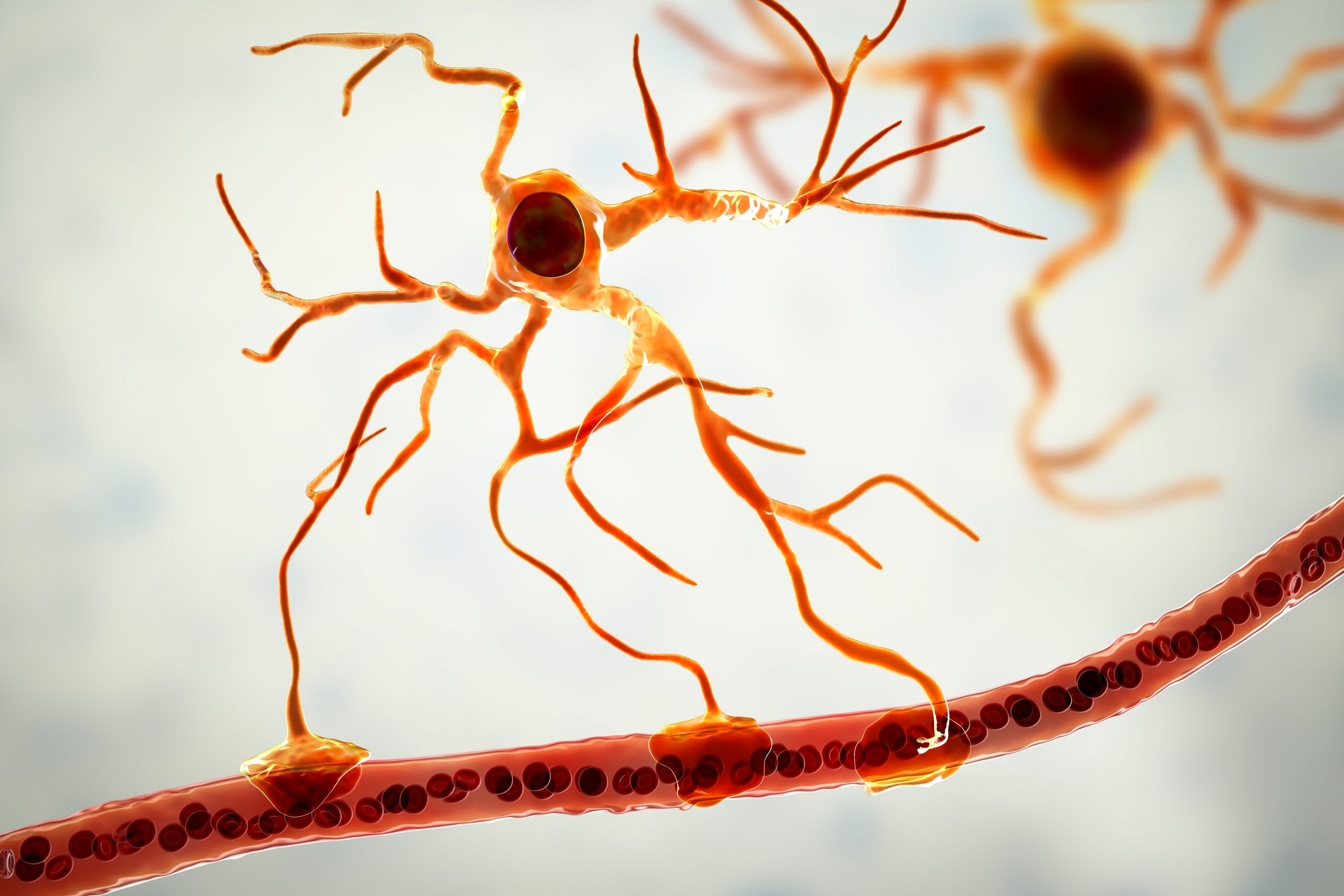


Astrocytes, the star-shaped cells that provide physical support for neurons in the brain, also appear to contribute to the development and progression of Parkinson’s disease, a recent study suggests.
Compared with healthy astrocytes, those containing Parkinson’s mutations showed multiple hallmarks of the disease, including the presence of alpha synuclein clumps, lower mitocondrial function, increased inflammatory responses, and changes in metabolism.
The research also shows that astrocytes derived from induced stem cells, which can be generated from a patient’s blood or skin cells, are useful models for better understanding the disease and investigating responses to potential therapies.
The study, “Metabolic alterations in Parkinson’s disease astrocytes,” was published in Scientific Reports.
Parkinson’s results from the death of neurons that produce dopamine, a neurotransmitter necessary to coordinate muscle movement. Loss of dopamine causes many of the disease’s symptoms, including tremors, loss of balance, and slowed movements.
While defects in dopaminergic neurons have been regarded as the main cause of Parkinson’s, a recent study also pointed to astrocytes as likely contributors in disease processes. Astrocytes are the most abundant cells in the brain; they provide physical support for neurons and help regulate the transmission of electrical impulses within the brain.
Since studies investigating the contribution of these cells to Parkinson’s are lacking, researchers at the University of Eastern Finland set out to examine patient-derived astrocytes and determine the changes in their cellular processes, compared with healthy astrocytes.
Because astrocytes cannot come directly from patients, induced pluripotent stem cells (iPSCs), which are generated from blood or skin cells, are seen as a helpful alternative. As their name suggests, these cells are pluripotent — meaning that, like other stem cells, they are able to differentiate into other cell types.
As such, researchers can collect blood or skin cells from a person with Parkinson’s and use these to generate iPSCs, which then can be differentiated into astrocytes. Since these astrocytes are derived from the iPSCs of a Parkinson’s patient, they will have the same genetic abnormalities, making them an apt model for study.
For this study, researchers generated astrocytes from two patients with familial Parkinson’s and two healthy controls. Both patients had a common mutation (the G2019S mutation) in the LRRK2 gene, the most commonly mutated gene associated with Parkinson’s. One patient also had a mutation in the GBA gene, a known risk factor for Parkinson’s.
The team created two cell lines from each individual; after six months in culture, all iPSC lines had generated comparable and functional astrocytes. An additional control cell line also was created. For that, iPSCs from the patient without the GBA mutation were corrected to carry a normal version of the LRRK2 gene.
Overall, patient-derived astrocytes produced significantly more alpha-synuclein than genetically corrected astrocytes or those derived from healthy people.
“It has been previously described that astrocytes accumulate neuron-derived α-synuclein as a mechanism of neuroprotection, [but] we found that in the absence of neurons the iPSC-derived PD [Parkinson’s disease] astrocytes themselves produced more α-synuclein [than] the control cells,” the researchers wrote.
These cells also were more reactive to inflammatory stimuli and more sensitive to inflammatory reactivation than control astrocytes, suggesting they may contribute to neuroinflammation in Parkinson’s.
They also had and had increased calcium levels. Calcium signaling is essential for nerve cell communication, but this signaling often is deregulated in the presence of alpha synuclein clumps and LRRK2 mutations.
Astrocytes with Parkinson’s mutations also showed lower mitochondrial function and fewer copies of mitochondrial DNA. These cells additionally had metabolic alterations compared to healthy astrocytes, particularly increased levels of polyamines and polyamine precursors and lower levels of the fat molecule lysophosphatidylethanolamine.
These mitochondrial and metabolic changes have been linked to Parkinson’s in prior studies, but researchers now demonstrated that astrocytes also manifest these alterations.
“While PD has been considered as a disease of the DA [dopaminergic] neurons, it is becoming more and more evident that other cell types of the CNS [central nervous system], including astrocytes, play an important role in PD pathogenesis,” the investigators wrote.
“The results provide evidence that LRRK2 and GBA mutant astrocytes are likely to contribute to PD progression and offer new perspectives for understanding the roles of astrocytes in the pathogenesis of PD,” Tuuli-Maria Sonninen, lead author of the study, said in a press release.