Surprising Link Between Parkinson’s and Gut’s Nervous System Found in Genome Study


Comparing the genomes of people with Parkinson’s disease to the specific genes expressed by cell types in the nervous system revealed an unexpected association between Parkinson’s and the digestive nervous system, according to a study.
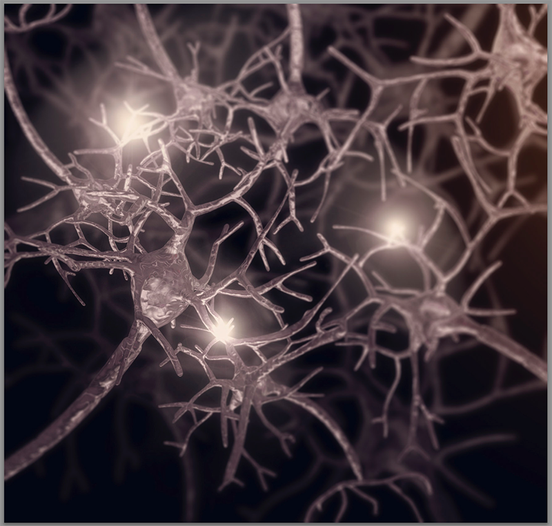
Another surprising result linked Parkinson’s to cells called oligodendrocytes, nervous system cells that produce a protein called myelin, which protectively coats other neurons to expedite the transmission of nerve signals.
The study, “Genetic identification of cell types underlying brain complex traits yields insights into the etiology of Parkinson’s disease,” was published in the journal Nature Genetics.
Parkinson’s disease is a nervous system disorder in which neurons in the substantia nigra region of the brain progressively die. Scientists believe that Parkinson’s may be caused by both genetic and environmental factors, though the exact root cause of the disease has not been identified.
To better understand the genetics of Parkinson’s, researchers at Karolinska Institutet in Sweden and the University of North Carolina used a technique called a genome-wide association study (GWAS) to identify variations in the genetic material of those with the disease.
GWAS compares genomic data from people with and without a condition to identify significant differences that may indicate the presence of that condition.
One downside of GWAS is that the data can be difficult to interpret. Genomic data can demonstrate only variations in genetic material; it cannot provide the context of how those variations occur in different cell types and tissues.
To address this shortcoming, the researchers paired GWAS genomic data with gene expression data to correlate genetic variations with specific cells and tissues.
They collected GWAS data for brain-specific conditions, including Parkinson’s, as well as conditions not specific to the brain, for verification.
These data were compared with gene expression profiles for 53 different human tissues from the Genotype-Tissue Expression Program database. The top 10% most specific genes were chosen to represent each tissue.
As expected, Parkinson’s was associated with the substantia nigra region of the brain, as well as the spinal cord.
This method also linked inflammatory bowel disease to the immune system and type 2 diabetes to the pancreas — expected results that verified the significance of their findings.
For more specific insight, the researchers then compared the GWAS data to cell-specific gene expression data from 39 unique cell types of the nervous system. Again, the top 10% most specific genes were chosen to represent each cell type.
These data were collected from mouse nervous cells, as the technique for collecting gene expression data is more accurate in mouse neurons than in human neurons. To account for this, the researchers did not include mouse genes that do not have a direct counterpart in humans.
This comparison identified four specific cell types: cholinergic neurons, monoaminergic neurons, the enteric (digestive) nervous system, and oligodendrocytes.
Cholinergic and monoaminergic neurons were expected results. Dopaminergic neurons — those that produce dopamine — are a type of monoaminergic neuron and are known to be affected by Parkinson’s.
The other two results were unexpected, though not without precedent.
With regards to the enteric nervous system, some research has shown that the digestive system may play a role in the development of Parkinson’s.
“As expected, we found that dopaminergic neurons were associated with Parkinson’s disease,” Patrick Sullivan, MD, one of the main study authors, said in a press release. “More surprisingly, we found that enteric neurons also seem to play an important role in the disorder, supporting the hypothesis that Parkinson’s disease starts in the gut.” Sullivan is a professor at both Karolinska Institutet and the University of North Carolina.
The association between Parkinson’s and oligodendrocytes also was an unexpected result. The researchers were able to verify this association in post-mortem human brains, finding that oligodendrocytes show varied genetic expression at even earlier stages of Parkinson’s than dopaminergic neurons do.
They suggest that oligodendrocytes may be a target for future Parkinson’s disease research.
“The fact that the animal studies pointed us to oligodendrocytes and that we were then able to show that these cells were also affected in patients suggests that the results may have clinical implications,” said Jens Hjerling-Leffler, PhD, a professor at the Karolinska Institutet and the study’s other main author.
Ultimately, by comparing genomic variance to specific gene expression data, the researchers demonstrated that enteric neurons and oligodendrocytes may play an important role in the progression of Parkinson’s disease.