Light-activated Protein Used to Improve Mitochondrial Function, Reduce Parkinson’s Symptoms in Fly Study


Using a light-activated protein to improve the function of mitochondria — cells’ powerhouses— could be a therapeutic strategy for Parkinson’s disease (PD), a new study done in flies suggests.
The results of the study, “Light-driven activation of mitochondrial proton-motive force improves motor behaviors in a Drosophila model of Parkinson’s disease,” were published in Communications Biology.
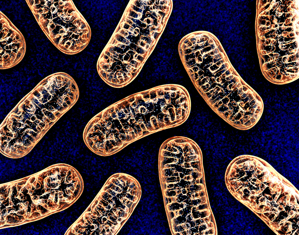
Mitochondria are organelles —specialized structures within a cell — that play a number of important biological roles within cells. Their most crucial function is generating adenosine triphosphate (ATP), which is the “energy currency” of the cell that fuels other cellular processes. The incorrect functioning of these organelles has been implicated in the development of Parkinson’s.
In particular, mutations in the gene that encodes the mitochondrial protein CHCHD2 — coiled-coil-helix-coiled-coil-helix domain containing 2 — have been shown to cause early-onset PD, as well as to impair the function of the mitochondria, specifically reducing the production of ATP. Flies lacking CHCHD2 also show PD-like symptoms, such as impaired motor abilities, aggregation or buildup of alpha-synuclein protein, and death of dopamine-producing (dopaminergic) neurons.
Normally, mitochondria create ATP using energy stored in the chemical bonds that are in the molecules of the food people eat. In the new study, the researchers created flies that got their energy a different way: from light.
Mitochondria generate ATP by moving hydrogen ions, or protons, across their membranes, which are their outer barrier layers. This creates a protonmotive force (delta p or Δp), which is what ultimately drives ATP generation.
The researchers created flies that had a protein called delta-rhodopsin (dR) on their mitochondrial membranes. dR is a protein originally found in archaea — single-celled organisms — that live in extremely high-salt conditions. This protein acts as a proton pump that basically can move protons across cellular membranes, such as the mitochondrial membrane. However, instead of getting its energy from food, dR moves protons using energy from particular wavelengths of light.
“Light-activated dR increases Δp, which promotes ATP production,” the researchers said.
The team tested whether light-activated dR could be used to rescue mitochondrial activity in flies lacking CHCHD2.
They found that, as expected, flies lacking CHCHD2 produced significantly less ATP than wild-type flies at 30 days of age. In flies with dR on their mitochondria, this also was the case when there was no dR-activating light. When that light was provided, however, the flies produced an amount of ATP that was not significantly different from the amount generated in wild-type flies.
Flies with light-activated dR also had lower levels of reactive oxygen species, highly reactive molecules that can damage cellular structures and are generated by the normal functioning of mitochondria. These flies also had lower levels of alpha-synuclein, a key protein that builds up in the brain in Parkinson’s disease. Flies with light-activated dR also had more surviving dopaminergic neurons — nerve cells that are progressively lost in Parkinson’s.
Additional experiments using a non-functional version of dR confirmed that these effects were a result of the protein itself, not some unforeseen non-specific effect of light on mitochondria.
“In conclusion,” the researchers said, “this study provides ‘proof of concept’ that Δp maintenance is beneficial for neuroprotection and that the development of proton pumps driven by optogenetic [light-driven] or pharmacogenetic [drug-driven] techniques is a potential therapeutic strategy for PD.”