Trial to Test New Imaging Agent for Visualizing Synapses in Living Brain
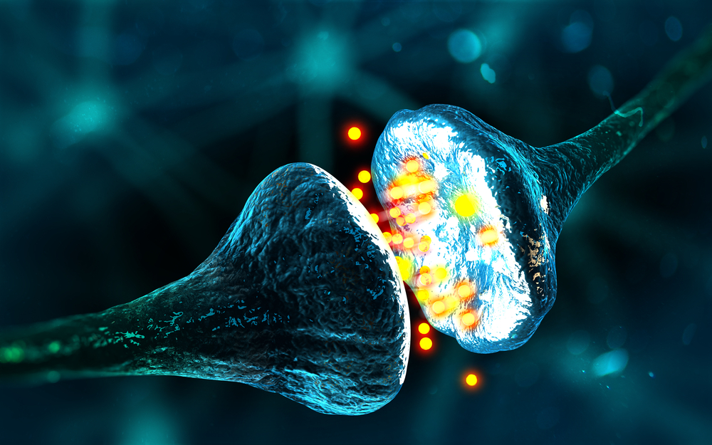


Rodin Therapeutics will evaluate a new imaging agent that allows the visualization of human synapses — the junctions between two nerve cells that allow them to communicate — in the living brain.
Results from the study will guide the company’s upcoming Phase 1b trial of a new therapeutic compound designed to strengthen and increase the number of synapses in patients with neurologic diseases, including Parkinson’s.
Several neurological and psychiatric diseases are characterized by defective synapses. However, there is currently no way to visualize synapses in the living brain. Tissues would need to be sampled, an invasive and unwanted procedure.
The new imaging agent, a radiotracer called [11C]UCB-J, targets the synaptic vesicle glycoprotein 2A (SV2A) and is used in positron emission tomography (PET). This protein regulates the release of neurotransmitters — chemical messengers used by the nervous system to transmit messages between neurons, or from neurons to muscles — at the synapse.
PET imaging uses small amounts of radioactive materials, called radiotracers, along with a special camera and computer to help evaluate organ and tissue function.
The trial, which will include both healthy volunteers and patients with Alzheimer’s disease, will produce brain scans following administration of the radiotracer. Participants will undergo scans at the beginning of the trial (baseline) and 28 days later.
The trial will determine the radiotracer’s robustness and potential usefulness in future trials.
“Most neurodegenerative disorders, including Alzheimer’s disease and Parkinson’s, are associated with deteriorating synapses — but until now, physicians and researchers have not been able to measure synaptic density in a living patient. This PET scan should allow us to visualize brain synaptic density in patients and possibly track their response to therapies over time,” J. Michael Ryan, MD, Rodin’s chief medical officer, said in a press release.
“Measuring synaptic density in a living human being holds tremendous potential for the diagnosis and treatment of a variety of neurologic diseases. We’re excited to be one of the first research centers to utilize this new imaging technology,” said Peter Paul De Deyn, MD, Ph.D., director of the Alzheimer’s Research Center Groningen, the Netherlands, one of the centers where the trial will take place.
“This tool has the potential to shape future clinical trials by offering an early signal about whether an investigational drug is driving molecular and structural changes in the brain,” said Philip Scheltens, MD, Ph.D., who directs the Alzheimer’s Center at VUmc, Amsterdam, another trial center. “We can then take the next step and assess whether those changes lead to functional and cognitive improvements in patients with neurodegenerative diseases.”