New Fluorescent Tools Help Researchers Track Protein Involved in Parkinson’s Disease
Written by |

Researchers at the University of Pennsylvania developed a strategy using fluorescence that allows them to track alpha-synuclein protein and follow its path inside neurons. This is important because while scientists know that alpha-synuclein protein is involved in neurodegenerative diseases like Parkinson’s, they aren’t exactly certain how the process functions.
These new monitoring tools could help scientists better understand neurodegenerative diseases and lead to progress on discovering new therapies or cures.
The study, “Selective imaging of internalized proteopathic α-synuclein seeds in primary neurons reveals mechanistic insight into transmission of synucleinopathies,” was published in the Journal of Biological Chemistry.
In Parkinson’s disease, the accumulation of misfolded alpha-synuclein protein forms fibrous deposits known as amyloid fibrils. These are the major components of Lewy bodies, the pathogenic, insoluble aggregates that develop inside neurons in patients with Parkinson’s disease.
Increasing evidence suggests that the misfolded proteins are transmitted directly from cell to cell, and this phenomenon underlies the disease’s progression. The mechanisms that allow these proteins to be transmitted, however, are not clearly understood. The changes the proteins cause once inside cells also have remained a mystery.
Now, the University of Pennsylvania research team has developed tools allowing them to track and quantify how the accumulation of misfolded alpha-synuclein is transmitted between neurons and what alterations happen inside the cell.
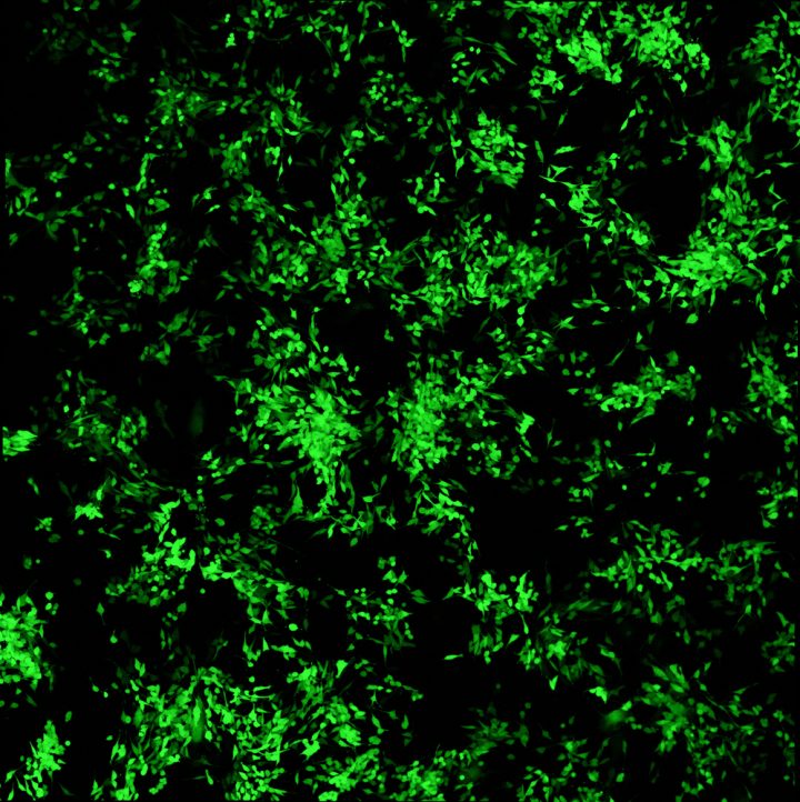
The team developed a simple method to observe alpha-synuclein fibrils entering cells. First, they produced alpha-synuclein fibrils tagged with fluorescent proteins so that by tracking the fluorescence, they could track the misfolded protein. Then, they added tagged alpha-synuclein to neurons in a culture.
Once together, a third element was added to the in vitro culture of neurons, a compound called trypan blue. This allows the “turning off” of fluorescent tags, and trypan blue cannot cross cell membranes.
So, when researchers added it, only the alpha-synuclein fibrils that had already entered neurons continued to glow. In all others, which remained locked outside the cells, the fluorescence was turned off.
To follow what happens to the fibrils once inside neurons, the team devised another strategy. Collaborating with the university’s chemistry department under the direction of E. James Petersson, the team built fibrils labeled with long-lasting fluorescent tags. These were further developed into two types that were sensitive or insensitive to acidity. Once again, by tracking the fluorescent fibrils, researchers could identify where the proteins ended up inside the cells.
The strategy revealed that fibrils were actively engulfed by the cell membrane and transported to waste disposal compartments called lysosomes. Fibrils remained in lysosomes for days.
“It’s amazing how much the cell is able to sequester,” Richard Karpowicz Jr., the study’s first author, said in a press release.
Some fibrils, however, were capable of escaping lysosomes and initiating protein aggregation. After inhibiting lysosome acidic activity with another chemical called chloroquine, researchers increased the number of alpha-synuclein protein ready to form aggregates. These results mirror the dysfunction of lysosomes often observed in patients with neurodegenerative diseases.
“We know that some of [the pathological proteins] somehow get out of the lysosomes,” said Virginia Lee, the study’s lead author. “But we don’t know how that happens.”
Overall, the findings represent a key strategy to monitor neurons’ uptake of alpha-synuclein misfolded protein. This will allow for rapid testing of pharmacological compounds targeting this process.
“Once you can look at [fibril] uptake into the cell, and quantify how much is inside the cell, then you can add small molecules to it to see if you can reduce uptake,” Lee said. “It’s really a simple assay and doesn’t take very long.”


